Yonsei Med J.
2009 Dec;50(6):845-847. 10.3349/ymj.2009.50.6.845.
A Case of Dysplastic Nevus of the External Auditory Canal Presenting with Conductive Hearing Loss
- Affiliations
-
- 1Department of Otorhinolaryngology-Head and Neck Surgery, Hallym University, Seoul, Korea. kcw5088@dreamwiz.com
- 2Department of Pathology, College of Medicine, Hallym University, Seoul, Korea.
- KMID: 1777101
- DOI: http://doi.org/10.3349/ymj.2009.50.6.845
Abstract
- A nevus which is a benign melanocytic neoplasm rarely occurs within the external auditory canal (EAC). A dysplastic nevus presents atypical features both clinically and histologically, and is important as a potential precursor for melanoma. We present a case of a 33-year-old female patient with a dysplastic nevus in her EAC. Physical examination revealed a protruding mass arising from the posterior wall of the left cartilaginous EAC. The mass showed clinically characteristic findings of a melanocytic nevus. The patient underwent excisional biopsy via a transcanal approach under local anesthesia. Histopathological examination revealed an intradermal nevus with atypical melanocytes without pleomorphism. There was no evidence of recurrence two years after surgical excision.
MeSH Terms
Figure
-

Fig. 1 Endoscopic image reveals a dome-shaped papular mass arising from the left cartilaginous ear canal's posterior wall.
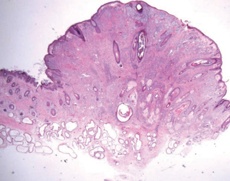
Fig. 2 Histopathologic findings of the mass showing nests of nevus cells in the dermis covered with normal skin (Hematoxylin-eosin, original magnification ×10).
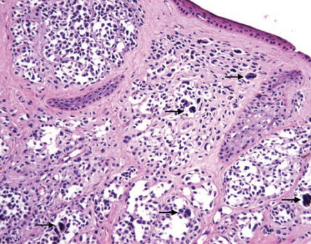
Fig. 3 Histopathologic findings of the mass showing scattered clusters which were composed of round to oval shaped melanocytic cells (black arrow) (Hematoxylin-eosin, original magnification ×200).
Reference
-
1. Lee FP. Pigmented nevus of the external auditory canal. Otolaryngol Head Neck Surg. 2006. 135:124–128.
Article2. Youngs R, Hawke M, Kwok P. Intradermal nevus of the ear canal. J Otolaryngol. 1988. 17:241–243.3. Deguine C, Pulec JL. Benign nevus of the external auditory canal. Ear Nose Throat J. 1998. 77:448.
Article4. Bothwell NE, Willard CC, Sorensen DM, Downey TJ. A rare case of a sebaceous nevus in the external auditory canal. Ear Nose Throat J. 2003. 82:38–41.
Article5. Kazikdas KC, Onal K, Kuehnel TS, Ozturk T. An intradermal nevus of the external auditory meatus. Eur Arch Otorhinolaryngol. 2006. 263:253–255.
Article6. Farrahi F, Egbert BM, Swetter SM. Histologic similarities between lentigo maligna and dysplastic nevus: importance of clinicopathologic distinction. J Cutan Pathol. 2005. 32:405–412.
Article7. Elder DE. Precursors to melanoma and their mimics: nevi of special sites. Mod Pathol. 2006. 19:S4–S20.
Article8. Naeyaert JM, Brochez L. Clinical practice. Dysplastic nevi. N Engl J Med. 2003. 349:2223–2240.9. Marghoob AA, Blum R, Nossa R, Busam KJ, Sachs D, Halpern A. Agminated atypical (dysplastic) nevi: case report and review of the literature. Arch Dermatol. 2001. 137:917–920.10. Tsao H, Bevona C, Goggins W, Quinn T. The transformation rate of moles (melanocytic nevi) into cutaneous melanoma: a population-based estimate. Arch Dermatol. 2003. 139:282–288.
Article
- Full Text Links
-
- Actions
-
Cited
- CITED
-
- Close
- Share
-
- Similar articles
-
- A Case of Intradermal Nevus of the External Auditory Canal
- A Case of Compound Nevus of the External Auditory Canal
- A Case of Langerhans Cell Histiocytosis involving the External Auditory Canal
- Compound Nevus Occurring Near External Auditory Canal: Successful Treatment by CO2 Laser Abrasion
- Irritated Seborrheic Keratosis of the External Auditory Canal which Caused Conductive Hearing Loss
