J Korean Med Sci.
2004 Oct;19(5):662-667. 10.3346/jkms.2004.19.5.662.
Predictive Value of Kushida Index and Acoustic Pharyngometry for the Evaluation of Upper Airway in Subjects With or Without Obstructive Sleep Apnea
- Affiliations
-
- 1Department of Otolaryngology-Head and Neck Surgery, St. Paul Hospital, The Catholic University Medical College, Seoul, Korea. dgjung@catholic.ac.kr
- 2Centre for Sleep Disorder and Respiratory Failure, Royal Prince Alfred Hospital, University of Sydney, NSW, Australia.
- KMID: 1733505
- DOI: http://doi.org/10.3346/jkms.2004.19.5.662
Abstract
- Acoustic pharyngometry is a relatively new noninvasive method that quantifies geometrically complexed pharyngeal dimensions. Our study aimed to investigate the predictability and usefulness of acoustic pharyngometry in diagnosis of obstructive sleep apnea (OSA), and we developed a prospective clinical trial in 16 subjects without apnea and 54 subjects with apnea. All seventy subjects received polysomnography (PSG) to assess the sleep architecture, including breathing and the degree of apnea hypopnea index. Acoustic pharyngometry was performed in four body positions (sitting, supine, right and left lateral) while awake with tidal breathing in addition to morphometric measurements (Kushida index) of oral cavity. This study shows that the cross-sectional area and volume of the upper airway is smaller in the supine position than any other positions. As well, the oropharyngeal junction area of the supine position is the most predictive parameter to discriminate between subjects with or without OSA. Acoustic pharyngometry can be a clinically useful tool for localizing the narrowed portion of the upper airway and predicting obstructive sleep apnea.
Keyword
MeSH Terms
Figure
-
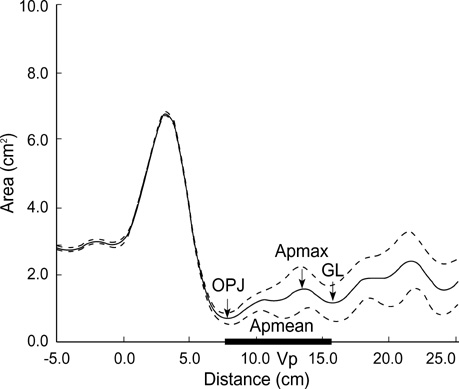
Fig. 1 Representative output of the acoustic pharyngometry. There are 5 parameters on graph as shown in arrow and thick line. OPJ, oropharyngeal junction area; Apmax, maximum pharyngeal area; GL, glottic area; Apmean, mean pharyngeal area from OPJ to GL; Vp, pharyngeal volume as the integrated area under the curve between the OPJ and the GL. All the parameters were calculated by computer system.
Reference
-
1. Young T, Palta M, Dempsey J, Skatrud J, Weber S, Badr S. The occurrence of sleep-disordered breathing among middle-aged adults. N Engl J Med. 1993. 328:1230–1235.
Article2. Grunstein RR, Hedner J, Grote L. Treatment options for sleep apnoea. Drugs. 2001. 61:237–251.
Article3. Schwab RJ, Gupta KB, Gefter WB, Metzger LJ, Hoffmann EA, Pack AI. Upper airway and soft tissue anatomy in normal subjects and patients with sleep-disordered breathing. Am J Respir Crit Care Med. 1995. 152:1673–1689.4. Kuna ST, Smickley JS. Superior pharyngeal constrictor activation in obstructive sleep apnea. Am J Respir Crit Care Med. 1997. 156:874–880.
Article5. Ikeda K, Oshima T, Shimomura A, Takasaka T. Surgical criteria for obstructive sleep apnea syndrome based on localization of upper airway collapse during sleep: a preliminary study. Tohoku J Exp Med. 1998. 185:1–8.
Article6. Shepard JW Jr, Gefter WB, Guilleminault C, Hoffman EA, Hoffstein V, Hudgel DW, Suratt PM, White DP. Evaluation of the upper airway in patients with obstructive sleep apnea. Sleep. 1991. 14:361–371.
Article7. Martin SE, Marshall I, Douglas NJ. The effect of posture on airway calibre in patients with the sleep apnoea/ hypopnoea syndrome. Am J Respir Crit Care Med. 1995. 152:721–724.8. Stanford W, Galvin J, Rooholamini M. Effects of awake tidal breathing, swallowing, nasal breathing, oral breathing and the Muller and Valsalva maneuvers on the dimensions of the upper airway: evaluation by ultrafast computerized tomography. Chest. 1988. 94:149–154.9. Brown IG, Zamel N, Hoffstein V. Pharyngeal cross-sectional area in normal men and women. J Appl Physiol. 1986. 61:890–895.
Article10. Sleep-related breathing disorders in adults: recommendations for syndrome definition and measurement techniques in clinical research. The Report of an American Academy of Sleep Medicine Task Force. Sleep. 1999. 22:667–689.11. Rubinstein I, McClean PA, Boucher R, Zamel N, Fredberg JJ, Hoffstein V. Effect of mouth piece, noseclips and head position on airway area measured by acoustic reflections. J Appl Physiol. 1987. 63:1469–1474.12. Jan MA, Marshall l, Douglas NJ. Effect of posture on upper airway dimensions in normal human. Am J Respir Crit Care Med. 1994. 149:145–148.
Article13. Kushida CA, Efron B, Guilleminault C. A predictive morphometric model for the obstructive sleep apnea syndrome. Ann Intern Med. 1997. 127:581–587.
Article14. Jackson AC, Butler JP, Millet EJ, Hoppin FG Jr, Dawson SV. Airway geometry by analysis of acoustic pulse response measurements. J Appl Physiol. 1977. 43:523–536.
Article15. Fredberg JJ, Wohl MEB, Glass GM, Dorkin HL. Airway area by acoustic reflections measured at the mouth. J Appl Physiol. 1980. 48:749–758.
Article16. D'Urzo AD, Rubinstein I, Lawson VG, Vassal KP, Rebuck AS, Slutsky AS, Hoffstein V. Comparison of glottic areas measured by acoustic reflections vs computerized tomography. J Appl Physiol. 1988. 64:367–370.17. Marshall I, Maran NJ, Martin S, Jan MA, Rimmington JE, Best JJ, Drummond GB, Douglas NJ. Acoustic reflectometry for airway measurements in man: implementation and validation. Physiol Meas. 1993. 14:157–169.
Article18. Malhotra A, Pillar G, Fogel R, Beauregard J, Edwards J, White DP. Upper-airway collapsibility: measurements and sleep effects. Chest. 2001. 120:156–161.
- Full Text Links
-
- Actions
-
Cited
- CITED
-
- Close
- Share
-
- Similar articles
-
- Predictability of Acoustic Pharyngometry for the Obstructive Sleep Apnea
- Acoustic Pharyngometry in Normal Adults
- Contemporary Methods of Upper Airway Evaluation in Obstructive Sleep Apnea Patients
- Upper airway studies in patients with obstructive sleep apnea syndrome
- Upper airway myofunctional exercise: a systematic review
