An Unusual Case with Membranous Lipodystrophy in a Hypertensive Patient with Transepidermal Elimination
- Affiliations
-
- 1Department of Dermatology, Yonsei University Wonju College of Medicine, Wonju, Korea. ahnsk@wonju.yonsei.ac.kr
- 2Department of Dermatology, Yonsei University College of Medicine, Seoul, Korea.
- KMID: 1715864
- DOI: http://doi.org/10.3349/ymj.2006.47.3.428
Abstract
- Membranous lipodystrophy represents a peculiar type of fat necrosis that is present in patients with various types of skin disease. It is characterized by the presence of microcysts and macrocysts and is lined by amorphous eosinophilic material with a crenelated arabesque appearance. These findings have been associated with lupus erythematosus, diabetes mellitus, erythema nodosum, trauma, etc. We report a case of a 43-year-old woman who had a red to purple asymptomatic indurated plaque, approximately seven cm in diameter and on the left arm. She was a chronic hepatitis B antigen carrier and had hypertension for four years. Histopathology of the biopsied lesion showed transepidermal elimination of altered collagen and elastic fibers, as well as membranous lipodystrophy changes. There were hypertensive vascular changes including lymphohistiocytic infiltration around the vascular wall, swelling of endothelial cells, increased thickness of the vascular walls, and narrowing of the lumen. We report a case showing transepidermal elimination with membranous lipodystrophy. We carefully suggest that the secondary phenomenon of transepidermal elimination was associated with membranous lipodystrophy and degenerate connective tissues.
MeSH Terms
Figure
-
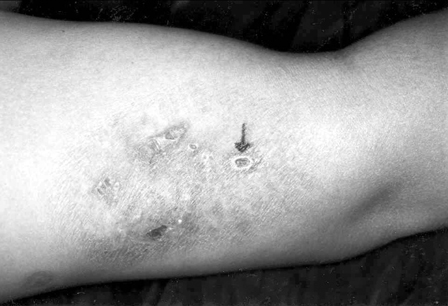
Fig. 1 The indurated plaque has multiple umbilicated papules with central keratin-filled plugs arranged in arcuate groups.
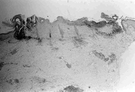
Fig. 2 An umbilical lesion shows a cup-shaped invagination of the epidermis that contains a large plug of keratin and degenerating nuclei of inflammatory cells (H&E, ×20).
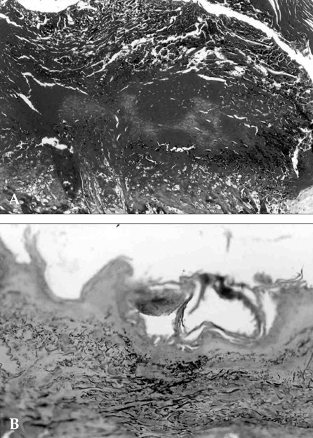
Fig. 3 (A) Degenerated collagen bundles are seen in the umbilicated lesion (Masson's trichrome, ×100). (B) Elastic fibers are seen in the invagination of the epidermis (Verhoeff-van Gieson, ×100).
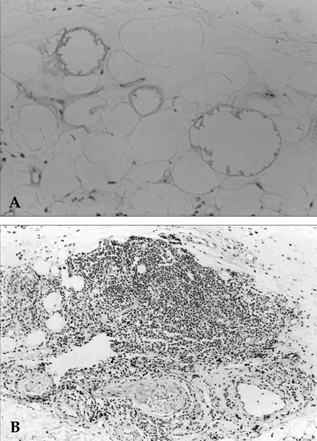
Fig. 4 (A) Lipomembranes have PAS positive feathery projections into the fat globules (PAS, ×200). (B) Blood vessels in the subcutis show infiltration of the vascular walls by lymphohistiocytes, swelling of endothelial cells and an increase the vascular walls' thickness (H&E, ×100).
Cited by 1 articles
-
Erythema Nodosum: Clinicopathologic Correlations and Their Use in Differential Diagnosis
Sang Won Yi, En Hyung Kim, Hee Young Kang, You Chan Kim, Eun-So Lee
Yonsei Med J. 2007;48(4):601-608. doi: 10.3349/ymj.2007.48.4.601.
Reference
-
1. Nasu T, Tsukahara Y, Terayama K. A lipid metabolic disease-"membranous lipodystrophy"-an autopsy case demonstrating numerous peculiar membrane-structures composed of compound lipid in bone and bone marrow and various adipose tissues. Acta Pathol Jpn. 1973. 23:539–558.2. Ahn SK, Yoo MS, Lee SH, Choi EH. A clinical and histopathological study of 22 patients with membranous lipodystrophy. Clin Exp Dermatol. 1996. 21:269–272.3. Patterson JW. The perforating disorders. J Am Acad Dermatol. 1984. 10:561–581.4. Rapini RP, Herbert AA, Drucker CR. Acquired perforating dermatosis. Evidence for combined transepidermal elimination of both collagen and elastic fibers. Arch Dermatol. 1989. 125:1074–1078.5. Machinami R. Membranous lipodystrophy-like changes in ischemic necrosis of the legs. Virchows Arch A Pathol Anat Histopathol. 1983. 399:191–205.6. Machinami R. Incidence of membranous lipodystrophy-like change among patients with limb necrosis caused by chronic arterial obstruction. Arch Pathol Lab Med. 1984. 108:823–826.7. Alegre VA, Winkelmann RK, Aliaga A. Lipomembranous changes in chronic panniculitis. J Am Acad Dermatol. 1988. 19:39–46.8. Diaz-Cascajo C, Borghi S. Subcutaneous pseudomembranous fat necrosis: New observations. J Cutan Pathol. 2002. 29:5–10.9. Korsgaard N, Aalkjaer C, Heagerty AM, Izzard AS, Mulvany MJ. Histology of subcutaneous small arteries from patients with essential hypertension. Hypertension. 1993. 22:523–526.10. Antonios TF, Singer DR, Markandu ND, Mortimer PS, MacGregor GA. Structural skin capillary rarefaction in essential hypertension. Hypertension. 1999. 33:998–1001.11. Alegre VA, Winkelmann RK, Aliaga A. Lipomembranous changes in chronic panniculitis. J Am Acad Dermatol. 1988. 19:39–46.
- Full Text Links
-
- Actions
-
Cited
- CITED
-
- Close
- Share
-
- Similar articles
-
- An Unusual Case of Transepidermal Elimination of Calcinosis Cutis
- A Case of Membranous Lipodystrophy with Stasis Dermatitis
- A Case of Membranous Lipodystrophy Observed in Lichen Amyloidosis
- A Case of Solitary Papular Angiokeratoma with Transepidermal Elimination
- A Case of Neurilemmoma Associated with Transepidermal Elimination
