Solitary Fibrous Tumor of the Pleura Manifesting as an Air-Containing Cystic Mass: Radiologic and Histopathologic Correlation
- Affiliations
-
- 1Department of Radiology, Seoul St. Mary's Hospital, College of Medicine, The Catholic University of Korea, Seoul 137-701, Korea. ami@catholic.ac.kr
- 2Department of Pathology, Seoul St. Mary's Hospital, College of Medicine, The Catholic University of Korea, Seoul 137-701, Korea.
- KMID: 1711468
- DOI: http://doi.org/10.3348/kjr.2013.14.6.981
Abstract
- Solitary fibrous tumor (SFT) is a rare mesenchymal neoplasm that typically presents as a well-defined lobular soft tissue mass commonly arising from the pleura. We report an extremely rare case of an SFT containing air arising from the right major fissure in a 58-year-old woman. Chest CT showed an ovoid air-containing cystic mass with an internal, homogeneously enhancing solid nodule. To our knowledge, this is the first case in the literature. The histopathologic findings were correlated with the radiologic findings, and the mechanism of air retention within the tumor is discussed.
MeSH Terms
Figure
-
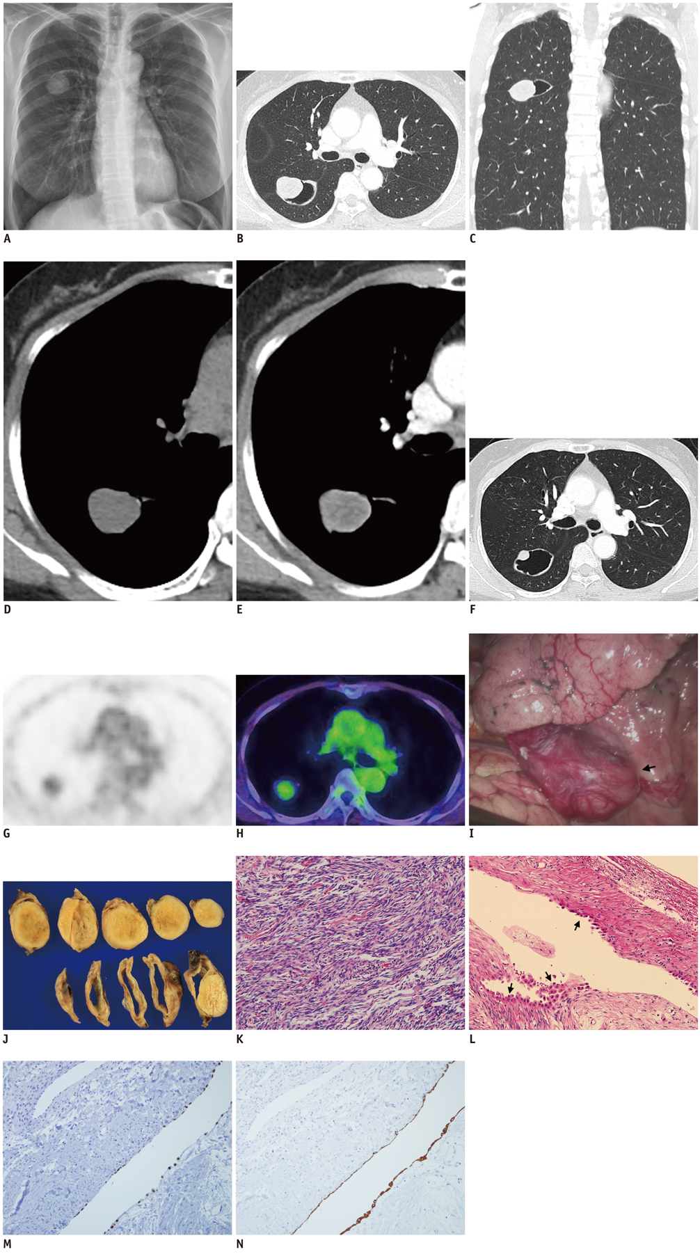
Fig. 1 Solitary fibrous tumor of pleura containing air in 58-year-old woman. A. Chest radiograph on admission shows air-containing cystic mass in right upper lung field. B-E. Chest CT images demonstrate 4.5 cm ovoid or spindle-shaped cystic mass along upper portion of right major fissure, which contains eccentrically located soft tissue nodule surrounded by large amount of air (B, C). Mass has irregular thin wall. Internal soft tissue nodule enhanced relatively homogeneously after contrast injection (D, E). F. Chest CT image obtained two years prior to current presentation reveals same-sized cystic mass, with much smaller internal solid nodule compared to recent CT images (B), suggesting nodule growth. G, H. F18-fluorodeoxyglucose (FDG) PET/CT scans of soft tissue nodule show mild FDG uptake with SUVmax of 2.3. I. Intraoperative photograph shows well-encapsulated pedunculated pleural tumor (arrow) arising from visceral pleura of right major fissure covering right upper lobe. J. Photograph of cut sections of resected specimen reveals well-encapsulated air-containing cystic mass with eccentrically located yellow-gray-colored solid nodule. K-N. Photomicrographs of specimen demonstrate variable components of spindle-shaped tumor cells and collagen fibers arranged in haphazard pattern, consistent with SFT in both internal nodular (K) and cystic (L) portions of tumor (hematoxylin-eosin stain, × 200). Cystic spaces are lined by cuboidal cells (arrows, L). Stains for TTF1 (M) and CK7 (N) show immunoreactivity in cystic lining epithelium, suggesting that they were pneumocytes.
Reference
-
1. Rosado-de-Christenson ML, Abbott GF, McAdams HP, Franks TJ, Galvin JR. From the archives of the AFIP: localized fibrous tumor of the pleura. Radiographics. 2003; 23:759–783.2. Mendelson DS, Meary E, Buy JN, Pigeau I, Kirschner PA. Localized fibrous pleural mesothelioma: CT findings. Clin Imaging. 1991; 15:105–108.3. Lee KS, Im JG, Choe KO, Kim CJ, Lee BH. CT findings in benign fibrous mesothelioma of the pleura: pathologic correlation in nine patients. AJR Am J Roentgenol. 1992; 158:983–986.4. Shim YS, Choi SJ, Kim HS, Lee JI. Solitary fibrous tumor of the trachea: CT findings with a pathological correlation. Korean J Radiol. 2008; 9:286–289.5. Cardillo G, Facciolo F, Cavazzana AO, Capece G, Gasparri R, Martelli M. Localized (solitary) fibrous tumors of the pleura: an analysis of 55 patients. Ann Thorac Surg. 2000; 70:1808–1812.6. England DM, Hochholzer L, McCarthy MJ. Localized benign and malignant fibrous tumors of the pleura. A clinicopathologic review of 223 cases. Am J Surg Pathol. 1989; 13:640–658.7. Bahk YW, Shinn KS, Choi BS. The air meniscus sign in sclerosing hemangioma of the lung. Radiology. 1978; 128:27–29.8. Nam JE, Ryu YH, Cho SH, Lee YJ, Kim HJ, Lee DY, et al. Air-trapping zone surrounding sclerosing hemangioma of the lung. J Comput Assist Tomogr. 2002; 26:358–361.9. Matsumoto K, Yamamoto T, Hisayoshi T, Asano G. Intravenous leiomyomatosis of the uterus with multiple pulmonary metastases associated with large bullae-like cyst formation. Pathol Int. 2001; 51:396–401.10. Wongsripuemtet J, Ruangchira-urai R, Stern EJ, Kanne JP, Muangman N. Benign metastasizing leiomyoma. J Thorac Imaging. 2012; 27:W41–W43.
- Full Text Links
-
- Actions
-
Cited
- CITED
-
- Close
- Share
-
- Similar articles
-
- A Case of Solitary Fibrous Tumor in the Cheek
- A Rare Solitary Fibrous Tumor of the Pleura with Extensive Cystic Change
- Spontaneous Interlobar Pneumothorax in a Localized Fibrous Tumor of in the Pleura
- Solitary Fibrous Tumor of the Adrenal Gland: A Case Report
- Ultrasonographic Localization of Solitary Fibrous Tumor of Pleura: Case Report
