Lab Med Online.
2014 Jul;4(3):152-156. 10.3343/lmo.2014.4.3.152.
Alterations of Complement C3 and C4 Levels in Delayed Testing
- Affiliations
-
- 1Department of Laboratory Medicine, Hanyang University Medical Center, Seoul, Korea. tykim@hanyang.ac.kr
- KMID: 1706852
- DOI: http://doi.org/10.3343/lmo.2014.4.3.152
Abstract
- BACKGROUND
In vitro levels of complement C3 and C4 proteins are sensitive to storage conditions. To avoid in vitro complement activation when testing is delayed, serum should be frozen at -20degrees C within 2 hr of venipuncture. However, this is impractical in routine laboratory work. Therefore, we investigated alterations in C3 and C4 levels in refrigerated specimens over time and derived formulae to estimate initial levels of complement concentrations in delayed testing.
METHODS
Ten fresh specimens were measured for C3 and C4 concentrations and were refrigerated at 4degrees C. We measured C3 and C4 levels in refrigerated samples daily for 4 days using an automated nephelometer (Beckman Coulter Inc., USA).
RESULTS
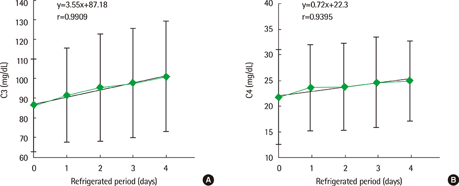
C3 and C4 levels were significantly increased over time in refrigerated specimens (P<0.001, P<0.001, respectively). The increments in C3 and C4 levels were described by the equations: C3 (mg/dL)=3.55x+87.18 (r=0.9909), and C4 (mg/dL)=0.72x+22.3 (r=0.9395), where x=the number of days samples were refrigerated before testing. Increases in C3 and C4 concentrations were described on a percentage basis by the equations: DeltaC3 (%)=4.14x+1.07 (r=0.9903), and DeltaC4 (%)=3.57x+2.48 (r=0.9405).
CONCLUSIONS
As the measured C3 and C4 concentrations increased by 3.55 mg/dL (4.1%) and 0.72 mg/dL (3.6%) per day in refrigerated specimens, the levels of C3 and C4 should be adjusted in delayed testing. We proposed that the formulae presented be used to back-calculate initial levels of C3 and C4 concentrations.
Keyword
MeSH Terms
Figure
Cited by 1 articles
-
Re-evaluation of the Utility of the CH50 Test Using Liposome Immunoassay
Jun Lee, La-He Jearn, Think-You Kim
Lab Med Online. 2021;11(3):171-176. doi: 10.47429/lmo.2021.11.3.171.
Reference
-
1. Massey HD, Richard AM. Mediators of inflammation: complement, cytokines, and adhesion molecules. In : Richard AM, Matthew RP, editors. Henry's clinical diagnosis and management by laboratory methods. 22nd ed. Philadelphia: Saunders Elsevier;2011. p. 914–932.2. Mollnes TE, Jokiranta TS, Truedsson L, Nilsson B, Rodriguez de Cordoba S, et al. Complement analysis in the 21st century. Mol Immunol. 2007; 44:3838–3849.
Article3. Ho A, Barr SG, Magder LS, Petri M. A decrease in complement is associated with increased renal and hematologic activity in patients with systemic lupus erythematosus. Arthritis Rheum. 2001; 44:2350–2357.
Article4. Illei GG, Takada K, Parkin D, Austin HA, Crane M, Yarboro CH, et al. Renal flares are common in patients with severe proliferative lupus nephritis treated with pulse immunosuppressive therapy: long-term follow up of a cohort of 145 patients participating in randomized controlled studies. Arthritis Rheum. 2002; 46:995–1002.
Article5. Okumura N, Nomura M, Tada T, Mitsuaki K, Takeshi M, Tsutomu K, et al. Effects of sample storage on serum C3 assay by immunonephelometry. Clin Lab Sci. 1990; 3:54–57.6. Mollnes TE, Garred P, Bergseth G. Effect of time, temperature and anticoagulants on in vitro complement activation: consequences for collection and preservation of samples to be examined for complement activation. Clin Exp Immunol. 1988; 73:484–488.7. Maguire OC, Curry MP, O'Gorman P, Parfrey N, Hegarty J, Cunningham SK. In vitro cold activation of complement shown by an overestimation of total complement 4: a study in patients with hepatitis C virus infection. Ann Clin Biochem. 2001; 38:687–693.
Article8. Thurman JM, Kulik L, Orth H, Wong M, Renner B, Sargsyan SA, et al. Detection of complement activation using monoclonal antibodies against C3d. J Clin Invest. 2013; 123:2218–2230.
Article9. Davies ET, Nasaruddin BA, Alhaq A, Senaldi G, Vergani D. Clinical application of new technique that measures C4d for assessment of activation of classical complement pathway. J Clin Pathol. 1988; 41:143–147.
Article10. David EN. Specimen collection and handling. In : Noel RR, Robert GH, editors. Manual of molecular and clinical laboratory immunology. 6th ed. Washington, DC: ASM Press;2002. p. 49–50.11. Pfeifer PH, Kawahara MS, Hugli TE. Possible mechanism for in vitro complement activation in blood and plasma samples: futhan/EDTA controls in vitro complement activation. Clin Chem. 1999; 45:1190–1199.
Article12. Newell S, Gorman JC, Bell E, Atkinson JP. Hemolytic and antigenic measurements of complement. A comparison of serum and plasma samples in normal individuals and patients. J Lab Clin Med. 1982; 100:437–444.
- Full Text Links
- Actions
-
Cited
- CITED
-
- Close
- Share
- Similar articles
-
- Re-evaluation of the Utility of the CH50 Test Using Liposome Immunoassay
- Fixation of Properdin and Factor B by Bullous Pemphigoid Antibody (in vitro Study)
- Changes of Immunoglobulin G , A , M and Complement C3 , C4 during Cardiopulmonary bypass under Fentanyl Anesthesia
- Pathology of C3 Glomerulonephritis
- Clinical significance of the immunological tests in rheumatoid arthritis


