Korean J Radiol.
2013 Apr;14(2):343-349. 10.3348/kjr.2013.14.2.343.
F-18 Fluorodeoxyglucose PET/CT and Post Hoc PET/MRI in a Case of Primary Meningeal Melanomatosis
- Affiliations
-
- 1Department of Nuclear Medicine, Dongnam Institute of Radiological & Medical Sciences (DIRAMS), Busan 619-953, Korea.
- 2Department of Nuclear Medicine, Kyungpook National University School of Medicine, Kyungpook National University Hospital, Daegu 700-721, Korea. abc2000@knu.ac.kr
- 3Department of Pathology, Kyungpook National University School of Medicine, Kyungpook National University Hospital, Daegu 700-721, Korea.
- 4Department of Neurosurgery, Kyungpook National University School of Medicine, Kyungpook National University Hospital, Daegu 700-721, Korea.
- 5Department of Nuclear Medicine, Samsung Medical Center, Sungkyunkwan University School of Medicine, Seoul 135-710, Korea.
- KMID: 1482798
- DOI: http://doi.org/10.3348/kjr.2013.14.2.343
Abstract
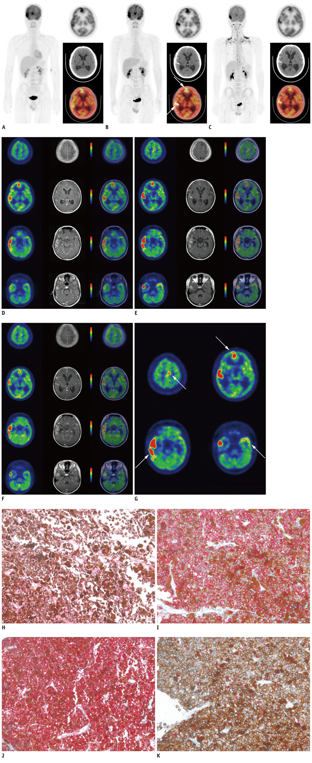
- Primary meningeal melanomatosis is a rare, aggressive variant of primary malignant melanoma of the central nervous system, which arises from melanocytes within the leptomeninges and carries a poor prognosis. We report a case of primary meningeal melanomatosis in a 17-year-old man, which was diagnosed with 18F-fluorodeoxyglucose (F-18 FDG) PET/CT, and post hoc F-18 FDG PET/MRI fusion images. Whole-body F-18 FDG PET/CT was helpful in ruling out the extracranial origin of melanoma lesions, and in assessing the therapeutic response. Post hoc PET/MRI fusion images facilitated the correlation between PET and MRI images and demonstrated the hypermetabolic lesions more accurately than the unenhanced PET/CT images. Whole body F-18 FDG PET/CT and post hoc PET/MRI images might help clinicians determine the best therapeutic strategy for patients with primary meningeal melanomatosis.
Keyword
MeSH Terms
-
Adolescent
Brain Neoplasms/*diagnosis/radionuclide imaging
Fluorodeoxyglucose F18/diagnostic use
Humans
*Magnetic Resonance Imaging
Male
Melanoma/*diagnosis/radionuclide imaging
Meningeal Neoplasms/*diagnosis/radionuclide imaging
*Positron-Emission Tomography and Computed Tomography
Radiopharmaceuticals/diagnostic use
Whole Body Imaging
Radiopharmaceuticals
Fluorodeoxyglucose F18
Figure
Reference
-
1. Savitz MH, Anderson PJ. Primary melanoma of the leptomeninges: a review. Mt Sinai J Med. 1974. 41:774–791.2. Smith AB, Rushing EJ, Smirniotopoulos JG. Pigmented lesions of the central nervous system: radiologic-pathologic correlation. Radiographics. 2009. 29:1503–1524.3. Gaetani P, Martelli A, Sessa F, Zappoli F, Rodriguez R, Baena . Diffuse leptomeningeal melanomatosis of the spinal cord: a case report. Acta Neurochir (Wien). 1993. 121:206–211.4. Sagiuchi T, Ishii K, Utsuki S, Asano Y, Tsukahara S, Kan S, et al. Increased uptake of technetium-99m-hexamethylpropyleneamine oxime related to primary leptomeningeal melanoma. AJNR Am J Neuroradiol. 2002. 23:1404–1406.5. Nicolaides P, Newton RW, Kelsey A. Primary malignant melanoma of meninges: atypical presentation of subacute meningitis. Pediatr Neurol. 1995. 12:172–174.6. Bang OY, Kim DI, Yoon SR, Choi IS. Idiopathic hypertrophic pachymeningeal lesions: correlation between clinical patterns and neuroimaging characteristics. Eur Neurol. 1998. 39:49–56.7. Zadro I, Brinar VV, Barun B, Ozretić D, Pazanin L, Grahovac G, et al. Primary diffuse meningeal melanomatosis. Neurologist. 2010. 16:117–119.8. Wadasadawala T, Trivedi S, Gupta T, Epari S, Jalali R. The diagnostic dilemma of primary central nervous system melanoma. J Clin Neurosci. 2010. 17:1014–1017.9. Pirini MG, Mascalchi M, Salvi F, Tassinari CA, Zanella L, Bacchini P, et al. Primary diffuse meningeal melanomatosis: radiologic-pathologic correlation. AJNR Am J Neuroradiol. 2003. 24:115–118.10. Bar-Shalom R, Yefremov N, Guralnik L, Gaitini D, Frenkel A, Kuten A, et al. Clinical performance of PET/CT in evaluation of cancer: additional value for diagnostic imaging and patient management. J Nucl Med. 2003. 44:1200–1209.11. Czernin J, Allen-Auerbach M, Schelbert HR. Improvements in cancer staging with PET/CT: literature-based evidence as of September 2006. J Nucl Med. 2007. 48:Suppl 1. 78S–88S.12. Louis DN, Ohgaki H, Wiestler OD, Cavenee WK, Burger PC, Jouvet A, et al. The 2007 WHO classification of tumours of the central nervous system. Acta Neuropathol. 2007. 114:97–109.13. Hayward RD. Malignant melanoma and the central nervous system. A guide for classification based on the clinical findings. J Neurol Neurosurg Psychiatry. 1976. 39:526–530.14. Nakhleh RE, Wick MR, Rocamora A, Swanson PE, Dehner LP. Morphologic diversity in malignant melanomas. Am J Clin Pathol. 1990. 93:731–740.15. Ohsie SJ, Sarantopoulos GP, Cochran AJ, Binder SW. Immunohistochemical characteristics of melanoma. J Cutan Pathol. 2008. 35:433–444.16. Brat DJ, Giannini C, Scheithauer BW, Burger PC. Primary melanocytic neoplasms of the central nervous systems. Am J Surg Pathol. 1999. 23:745–754.17. Liubinas SV, Maartens N, Drummond KJ. Primary melanocytic neoplasms of the central nervous system. J Clin Neurosci. 2010. 17:1227–1232.18. Lee NK, Lee BH, Hwang YJ, Sohn MJ, Chang S, Kim YH, et al. Findings from CT, MRI, and PET/CT of a primary malignant melanoma arising in a spinal nerve root. Eur Spine J. 2010. 19:Suppl 2. S174–S178.19. Bastiaannet E, Oyen WJ, Meijer S, Hoekstra OS, Wobbes T, Jager PL, et al. Impact of [18F]fluorodeoxyglucose positron emission tomography on surgical management of melanoma patients. Br J Surg. 2006. 93:243–249.
- Full Text Links
- Actions
-
Cited
- CITED
-
- Close
- Share
- Similar articles
-
- Use of 18F-FDG PET/CT in Second Primary Cancer
- Multifocal Head and Neck Paraganglioma Evaluated with Different PET Tracers: Comparison Between Fluorine-18-Fluorodeoxyglucose and Gallium-68-Somatostatin Receptor PET/CT
- Fluorodeoxyglucose Positron-Emission Tomography/Computed Tomography and Magnetic Resonance Imaging for Adverse Local Tissue Reactions near Metal Implants after Total Hip Arthroplasty: A Preliminary Report
- The Use of PET in Esophageal Cancer
- CT, MRI, and 18F-Fluorodeoxyglucose Positron Emission Tomography/CT Features of Primary Mucosal Melanoma Involving the Lacrimal Drainage Apparatus: A Case Report


