Skeletal and dentoalveolar changes after miniscrew-assisted rapid palatal expansion in young adults: A cone-beam computed tomography study
- Affiliations
-
- 1Department of Orthodontics, Institute of Craniofacial Deformity, College of Dentistry, Yonsei University, Seoul, Korea. yoonjchoi@yuhs.ac
- 2Graduate of Harvard School of Dental Medicine, Harvard University, Boston, MA, USA.
- KMID: 2400385
- DOI: http://doi.org/10.4041/kjod.2017.47.2.77
Abstract
OBJECTIVE
The aim of this study was to evaluate the skeletal and dentoalveolar changes after miniscrew-assisted rapid palatal expansion (MARPE) in young adults by cone-beam computed tomography (CBCT).
METHODS
This retrospective study included 14 patients (mean age, 20.1 years; range, 16-26 years) with maxillary transverse deficiency treated with MARPE. Skeletal and dentoalveolar changes were evaluated using CBCT images acquired before and after expansion. Statistical analyses were performed using paired t-test or Wilcoxon signed-rank test according to normality of the data.
RESULTS
The midpalatal suture was separated, and the maxilla exhibited statistically significant lateral movement (p < 0.05) after MARPE. Some of the landmarks had shifted forwards or upwards by a clinically irrelevant distance of less than 1 mm. The amount of expansion decreased in the superior direction, with values of 5.5, 3.2, 2.0, and 0.8 mm at the crown, cementoenamel junction, maxillary basal bone, and zygomatic arch levels, respectively (p < 0.05). The buccal bone thickness and height of the alveolar crest had decreased by 0.6-1.1 mm and 1.7-2.2 mm, respectively, with the premolars and molars exhibiting buccal tipping of 1.1°-2.9°.
CONCLUSIONS
Our results indicate that MARPE is an effective method for the correction of maxillary transverse deficiency without surgery in young adults.
Keyword
MeSH Terms
Figure
-
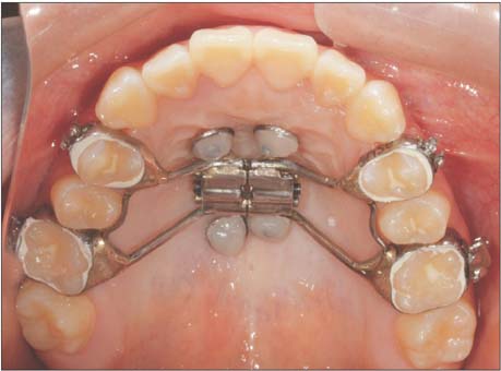
Figure 1 Clinical application of miniscrew-assisted rapid palatal expansion.
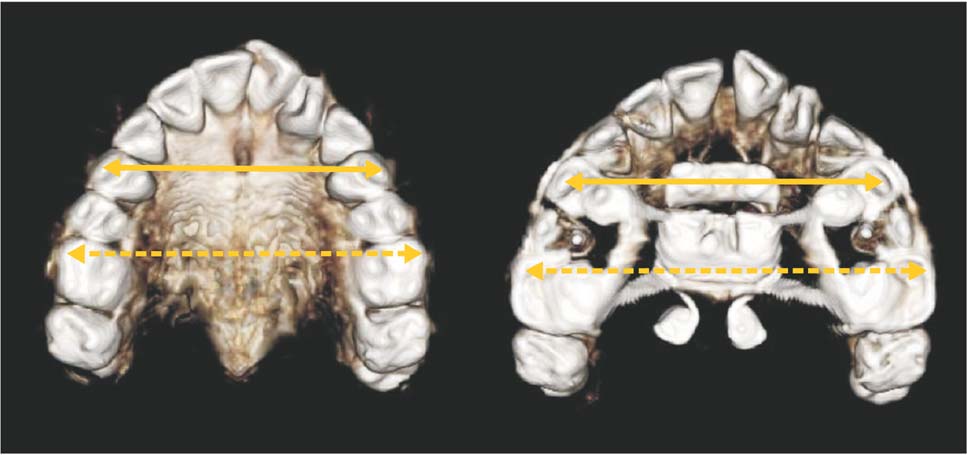
Figure 2 Three-dimensional tooth models used for the cone-beam computed tomography assessment of interpremolar and intermolar widths after miniscrew-assisted rapid palatal expansion. Solid arrow, interpremolar width; dashed arrow, intermolar width.

Figure 3 Two-dimensional posteroanterior cephalogram reconstructed from a three-dimensional skull model. Refer to Table 1 for the definitions of abbreviations.
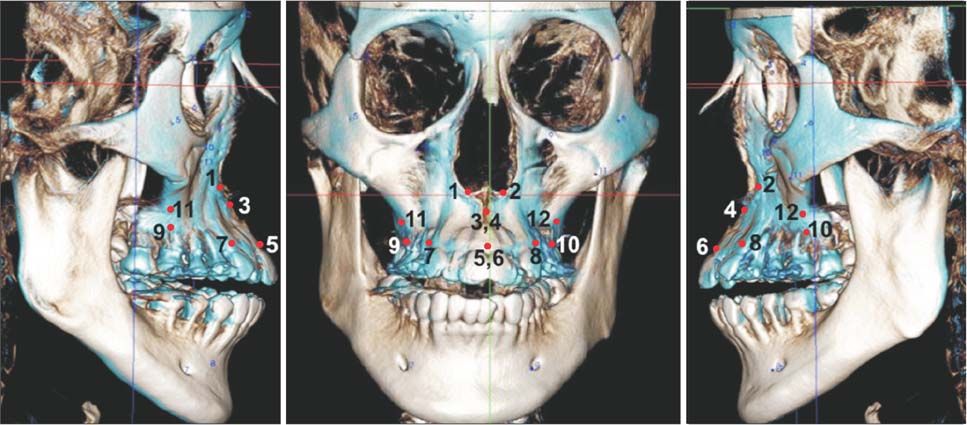
Figure 4 Superimposition of three-dimensional cone-beam computed tomography images acquired before (white) and after (blue) miniscrew-assisted rapid palatal expansion. 1 and 2, alare, right and left; 3 and 4, A-point, right and left; 5 and 6, prosthion, right and left; 7 and 8, ectocanine, right and left; 9 and 10, ectomolare, right and left; 11 and 12, processus zygomaticus, right and left.

Figure 5 Coronal cone-beam computed tomography images acquired before expansion at furcations of the first premolar (left) and first molar (right). a, buccal bone thickness; b, buccal alveolar height. Buccal bone thickness and alveolar height were measured on the right and left sides, and the mean value of the two measurements was calculated.
Cited by 9 articles
-
Stability of bimaxillary surgery involving intraoral vertical ramus osteotomy with or without presurgical miniscrew-assisted rapid palatal expansion in adult patients with skeletal Class III malocclusion
Yoon-Soo Ahn, Sung-Hwan Choi, Kee-Joon Lee, Young-Soo Jung, Hyoung-Seon Baik, Hyung-Seog Yu
Korean J Orthod. 2020;50(5):304-313. doi: 10.4041/kjod.2020.50.5.304.Stability of dental, alveolar, and skeletal changes after miniscrew-assisted rapid palatal expansion
Hyun-Mook Lim, Young-Chel Park, Kee-Joon Lee, Kyung-Ho Kim, Yoon Jeong Choi
Korean J Orthod. 2017;47(5):313-322. doi: 10.4041/kjod.2017.47.5.313.Investigation of the effects of miniscrew-assisted rapid palatal expansion on airflow in the upper airway of an adult patient with obstructive sleep apnea syndrome using computational fluid-structure interaction analysis
Jae-Sik Hur, Hyoung-Ho Kim, Jin-Young Choi, Sang-Ho Suh, Seung-Hak Baek
Korean J Orthod. 2017;47(6):353-364. doi: 10.4041/kjod.2017.47.6.353.Investigation of the effects of miniscrew-assisted rapid palatal expansion on airflow in the upper airway of an adult patient with obstructive sleep apnea syndrome using computational fluid-structure interaction analysis
Jae-Sik Hur, Hyoung-Ho Kim, Jin-Young Choi, Sang-Ho Suh, Seung-Hak Baek
Korean J Orthod. 2017;47(6):353-364. doi: 10.4041/kjod.2017.47.6.353.Dentofacial transverse development in Koreans according to skeletal maturation: A cross-sectional study
Soonshin Hwang, Yoonjeong Noh, Yoon Jeong Choi, Chooryung Chung, Hye Sun Lee, Kyung-Ho Kim
Korean J Orthod. 2018;48(1):39-47. doi: 10.4041/kjod.2018.48.1.39.Predictors of midpalatal suture expansion by miniscrew-assisted rapid palatal expansion in young adults: A preliminary study
Hyerin Shin, Chung-Ju Hwang, Kee-Joon Lee, Yoon Jeong Choi, Sang-Sun Han, Hyung Seog Yu
Korean J Orthod. 2019;49(6):360-371. doi: 10.4041/kjod.2019.49.6.360.Short-term impact of microimplant-assisted rapid palatal expansion on the nasal soft tissues in adults: A three-dimensional stereophotogrammetry study
Seung-Ryeol Lee, Jin-woo Lee, Dong-Hwa Chung, Sang-min Lee
Korean J Orthod. 2020;50(2):75-85. doi: 10.4041/kjod.2020.50.2.75.Effectiveness of miniscrew assisted rapid palatal expansion using cone beam computed tomography: A systematic review and meta-analysis
Patchaya Siddhisaributr, Kornkanok Khlongwanitchakul, Niwat Anuwongnukroh, Somchai Manopatanakul, Nita Viwattanatipa
Korean J Orthod. 2022;52(3):182-200. doi: 10.4041/kjod21.256.Skeletal and dentoalveolar effects of different types of microimplant-assisted rapid palatal expansion
Hyeong-Yoon Choi, Sang-Min Lee, Jin-Woo Lee, Dong-Hwa Chung, Mo-Hyeon Lee
Korean J Orthod. 2023;53(4):241-253. doi: 10.4041/kjod23.036.
Reference
-
1. da Silva Filho OG, Montes LA, Torelly LF. Rapid maxillary expansion in the deciduous and mixed dentition evaluated through posteroanterior cephalometric analysis. Am J Orthod Dentofacial Orthop. 1995; 107:268–275.
Article2. Haas AJ. Palatal expansion: just the beginning of dentofacial orthopedics. Am J Orthod. 1970; 57:219–255.
Article3. Shetty V, Caridad JM, Caputo AA, Chaconas SJ. Biomechanical rationale for surgical-orthodontic expansion of the adult maxilla. J Oral Maxillofac Surg. 1994; 52:742–749. discussion 750-1.
Article4. Asscherickx K, Govaerts E, Aerts J, Vande Vannet B. Maxillary changes with bone-borne surgically assisted rapid palatal expansion: A prospective study. Am J Orthod Dentofacial Orthop. 2016; 149:374–383.
Article5. Williams BJ, Currimbhoy S, Silva A, O'Ryan FS. Complications following surgically assisted rapid palatal expansion: a retrospective cohort study. J Oral Maxillofac Surg. 2012; 70:2394–2402.
Article6. Persson M, Thilander B. Palatal suture closure in man from 15 to 35 years of age. Am J Orthod. 1977; 72:42–52.
Article7. Stuart DA, Wiltshire WA. Rapid palatal expansion in the young adult: time for a paradigm shift? J Can Dent Assoc. 2003; 69:374–377.8. Rungcharassaeng K, Caruso JM, Kan JY, Kim J, Taylor G. Factors affecting buccal bone changes of maxillary posterior teeth after rapid maxillary expansion. Am J Orthod Dentofacial Orthop. 2007; 132:428.e1–428.e8.
Article9. Capelozza Filho L, Cardoso Neto J, da Silva Filho OG, Ursi WJ. Non-surgically assisted rapid maxillary expansion in adults. Int J Adult Orthodon Orthognath Surg. 1996; 11:57–66. discussion 67-70.10. Thilander B, Nyman S, Karring T, Magnusson I. Bone regeneration in alveolar bone dehiscences related to orthodontic tooth movements. Eur J Orthod. 1983; 5:105–114.
Article11. Ramieri GA, Spada MC, Austa M, Bianchi SD, Berrone S. Transverse maxillary distraction with a bone-anchored appliance: dento-periodontal effects and clinical and radiological results. Int J Oral Maxillofac Surg. 2005; 34:357–363.
Article12. Lee KJ, Park YC, Park JY, Hwang WS. Miniscrew-assisted nonsurgical palatal expansion before orthognathic surgery for a patient with severe mandibular prognathism. Am J Orthod Dentofacial Orthop. 2010; 137:830–839.
Article13. Mah JK, Danforth RA, Bumann A, Hatcher D. Radiation absorbed in maxillofacial imaging with a new dental computed tomography device. Oral Surg Oral Med Oral Pathol Oral Radiol Endod. 2003; 96:508–513.
Article14. Akyalcin S, Schaefer JS, English JD, Stephens CR, Winkelmann S. A cone-beam computed tomography evaluation of buccal bone thickness following maxillary expansion. Imaging Sci Dent. 2013; 43:85–90.
Article15. Christie KF, Boucher N, Chung CH. Effects of bonded rapid palatal expansion on the transverse dimensions of the maxilla: a cone-beam computed tomography study. Am J Orthod Dentofacial Orthop. 2010; 137:4 Suppl. S79–S85.
Article16. Kartalian A, Gohl E, Adamian M, Enciso R. Cone-beam computerized tomography evaluation of the maxillary dentoskeletal complex after rapid palatal expansion. Am J Orthod Dentofacial Orthop. 2010; 138:486–492.
Article17. Timock AM, Cook V, McDonald T, Leo MC, Crowe J, Benninger BL, et al. Accuracy and reliability of buccal bone height and thickness measurements from cone-beam computed tomography imaging. Am J Orthod Dentofacial Orthop. 2011; 140:734–744.
Article18. Vanarsdall RL Jr. Transverse dimension and long-term stability. Semin Orthod. 1999; 5:171–180.
Article19. Magnusson A, Bjerklin K, Kim H, Nilsson P, Marcusson A. Three-dimensional assessment of transverse skeletal changes after surgically assisted rapid maxillary expansion and orthodontic treatment: a prospective computerized tomography study. Am J Orthod Dentofacial Orthop. 2012; 142:825–833.
Article20. Corbridge JK, Campbell PM, Taylor R, Ceen RF, Buschang PH. Transverse dentoalveolar changes after slow maxillary expansion. Am J Orthod Dentofacial Orthop. 2011; 140:317–325.
Article21. Garib DG, Henriques JF, Janson G, de Freitas MR, Fernandes AY. Periodontal effects of rapid maxillary expansion with tooth-tissue-borne and tooth-borne expanders: a computed tomography evaluation. Am J Orthod Dentofacial Orthop. 2006; 129:749–758.
Article22. Jung PK, Lee GC, Moon CH. Comparison of cone-beam computed tomography cephalometric measurements using a midsagittal projection and conventional two-dimensional cephalometric measurements. Korean J Orthod. 2015; 45:282–288.
Article23. Melsen B. Palatal growth studied on human autopsy material. A histologic microradiographic study. Am J Orthod. 1975; 68:42–54.24. El H, Palomo JM. Three-dimensional evaluation of upper airway following rapid maxillary expansion: a CBCT study. Angle Orthod. 2014; 84:265–273.
Article25. Pinto PX, Mommaerts MY, Wreakes G, Jacobs WV. Immediate postexpansion changes following the use of the transpalatal distractor. J Oral Maxillofac Surg. 2001; 59:994–1000. discussion 1001.
Article26. Bazargani F, Feldmann I, Bondemark L. Three-dimensional analysis of effects of rapid maxillary expansion on facial sutures and bones. Angle Orthod. 2013; 83:1074–1082.
Article27. Lione R, Franchi L, Cozza P. Does rapid maxillary expansion induce adverse effects in growing subjects? Angle Orthod. 2013; 83:172–182.
Article28. Gurgel JA, Tiago CM, Normando D. Transverse changes after surgically assisted rapid palatal expansion. Int J Oral Maxillofac Surg. 2014; 43:316–322.
Article29. Park HS, Lee YJ, Jeong SH, Kwon TG. Density of the alveolar and basal bones of the maxilla and the mandible. Am J Orthod Dentofacial Orthop. 2008; 133:30–37.
Article30. Tian YL, Liu F, Sun HJ, Lv P, Cao YM, Yu M, Yue Y. Alveolar bone thickness around maxillary central incisors of different inclination assessed with cone-beam computed tomography. Korean J Orthod. 2015; 45:245–252.
Article31. Baysal A, Uysal T, Veli I, Ozer T, Karadede I, Hekimoglu S. Evaluation of alveolar bone loss following rapid maxillary expansion using cone-beam computed tomography. Korean J Orthod. 2013; 43:83–95.
Article32. Handelman CS, Wang L, BeGole EA, Haas AJ. Nonsurgical rapid maxillary expansion in adults: report on 47 cases using the Haas expander. Angle Orthod. 2000; 70:129–144.
- Full Text Links
-
- Actions
-
Cited
- CITED
-
- Close
- Share
-
- Similar articles
-
- Skeletal and dentoalveolar changes after miniscrew-assisted rapid palatal expansion in young adults: A cone-beam computed tomography study
- Stability of dental, alveolar, and skeletal changes after miniscrew-assisted rapid palatal expansion
- Skeletal and dentoalveolar effects of different types of microimplant-assisted rapid palatal expansion
- Predictors of midpalatal suture expansion by miniscrew-assisted rapid palatal expansion in young adults: A preliminary study
- Effectiveness of miniscrew assisted rapid palatal expansion using cone beam computed tomography: A systematic review and meta-analysis
