Mid-Ventricular Obstructive Hypertrophic Cardiomyopathy Associated with an Apical Aneurysm: Evaluation of Possible Causes of Aneurysm Formation
- Affiliations
-
- 1Department of Cardiology, Nihon University School of Medicine, Tokyo, Japan. yuichis@med.nihon-u.ac.jp
- 2Department of Cardiovascular and Respiratory Medicine, Shiga University of Medical Science, Seta Otsu, Shiga, Japan.
- KMID: 1122630
- DOI: http://doi.org/10.3349/ymj.2007.48.5.879
Abstract
- Mid-ventricular obstructive hypertrophic cardiomyopathy (MVOHCM) is a rare type of cardiomyopathy, associated with apical aneurysm formation in some cases. We report a patient presenting with ventricular fibrillation, an ECG with an above normal ST segment, and elevated levels of cardiac enzymes but normal coronary arteries. Left ventriculography revealed a left ventricular obstruction without apical aneurysm. There was a significant pressure gradient between the apical and basal sites of the left ventricle. Cine magnetic resonance imaging (MRI), performed on the 10th hospital day, showed asymmetric septal hypertrophy, mid-ventricular obstruction, and an apical aneurysm with a thrombus. The first evaluation by contrast-enhanced imaging showed a subendocardial perfusion defect and delayed enhancement. It was speculated that the intraventricular pressure gradient, due to mid- ventricular obstruction, triggered myocardial infarction, which subsequently resulted in apical aneurysm formation.
MeSH Terms
Figure
-
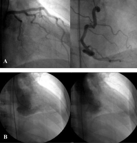
Fig. 1 (A) Coronary aniogram showing normal left (left panel) and right (right panel) coronary arteries. (B) Left ventriculogram in the 30° right anterior oblique projection, showing a small left ventricular cavity during diastole (left panel) and complete obstruction of the apical site during systole (right panel).
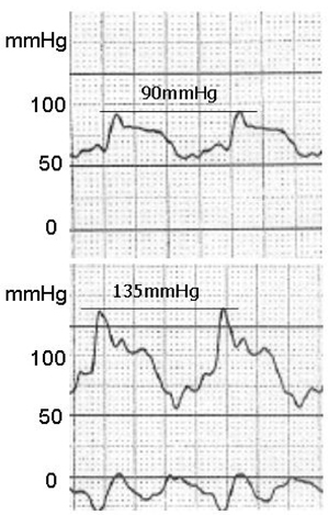
Fig. 2 Left ventricular pressure in the apex (upper panel) and in the base (lower panel), showing a pressure gradient of 45mmHg
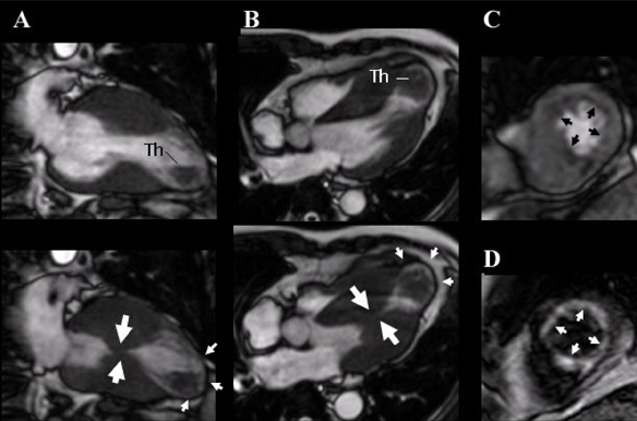
Fig. 3 ECG-gated, cine magnetic resonance imaging of the long axis (A) and 4-chamber view (B), showing mid-ventricular obstruction during systole (large arrows) (lower panels), an apical aneurysm (small arrows), and a thrombus (Th, upper panels) during diastole. Contrast-enhanced first-pass myocardial perfusion (C) and delayed enhancement (D) images, showing a subendomyocardial perfusion defect (arrows) and delayed enhancement (arrows), respectively.
Reference
-
1. Akutsu Y, Shinozuka A, Huang TY, Watanabe T, Yamada T, Yamanaka H, et al. Hypertrophic cardiomyopathy with apical left ventricular aneurysm. Jpn Circ J. 1988. 62:127–131.
Article2. Lin CS, Chen CH, Ding PY. Apical hypertrophic cardiomyopathy mimicking acute myocardial infarction. Int J Cardiol. 1998. 64:305–307.
Article3. Harada K, Shimizu T, Sugishita Y, Yao A, Suzuki J, Takenaka K, et al. Hypertrophic cardiomyopathy with midventricular obstruction and apical aneurysm: a case report. Jpn Circ J. 2001. 65:915–919.
Article4. Ito N, Suzuki M, Enjoji Y, Nakamura M, Namiki A, Hase H, et al. Hypertrophic cardiomyopathy with mid-ventricular obstruction complicated with apical left ventricular aneurysm and ventricular tachycardia: a case report. J Cardiol. 2002. 39:213–219.5. Tse HF, Ho HH. Sudden cardiac death caused by hypertrophic cardiomyopathy associated with midventricular obstruction and apical aneurysm. Heart. 2003. 89:178.
Article6. Tengiz I, Ercan E, Türk UO. Percutaneous myocardial ablation for left mid-ventricular obstructive hypertrophic cardiomyopathy. Int J Cardiovasc Imaging. 2006. 22:13–18.
Article7. Matsubara K, Nakamura T, Kuribayashi T, Azuma A, Nakagawa M. Sustained cavity obliteration and apical aneurysm formation in apical hypertrophic cardiomyopathy. J Am Coll Cardiol. 2003. 42:288–295.
Article8. Maron BJ, Hauser RG, Roberts WC. Hypertrophic cardiomyopathy with left ventricular apical diverticulum. Am J Cardiol. 1999. 77:1263–1265.
Article9. Ward RP, Pokharna HK, Lang RM, Williams KA. Resting "Solar Polar" map pattern and reduced apical flow reserve: characteristics of apical hypertrophic cardiomyopathy on SPECT myocardial perfusion imaging. J Nucl Cardiol. 2003. 10:506–512.
Article10. Fighali S, Krajcer Z, Edelman S, Leachman RD. Progression of hypertrophic cardiomyopathy into a hypokinetic left ventricle: higher incidence in patients with midventricular obstruction. J Am Coll Cardiol. 1987. 9:288–294.
Article11. Nakamura T, Matsubara K, Furukawa K, Azuma A, Sugihara H, Katsume H, et al. Diastolic paradoxic jet flow in patients with hypertrophic cardiomyopathy: evidence of concealed apical asynergy with cavity obliteration. J Am Coll Cardiol. 1992. 19:516–524.
Article12. Maron BJ, Casey SA, Poliac LC, Gohman TE, Almquist AK, Aeppli DM. Clinical course of hypertrophic cardiomyopathy in a regional United States cohort. JAMA. 1999. 281:650–655.
Article13. Maron BJ, Nishimura RA, McKenna WJ, Rakowski H, Josephson ME, Kieval RS. Assessment of permanent dual-chamber pacing as a treatment for drug-refractory symptomatic patients with obstructive hypertrophic cardiomyopathy. A randomized, double-blind, crossover study (M-PATHY). Circulation. 1999. 99:2927–2933.
Article14. Watanabe H, Kibira S, Saito T, Shimizu H, Abe T, Nakajima I, et al. Beneficial effect of dual-chamber pacing for a left mid-ventricular obstruction with apical aneurysm. Circ J. 2002. 66:981–984.
Article15. Seggewiss H, Faber L. Percutaneous septal ablation for hypertrophic cardiomyopathy and mid-ventricular obstruction. Eur J Echocardiogr. 2000. 1:277–280.
Article
- Full Text Links
-
- Actions
-
Cited
- CITED
-
- Close
- Share
-
- Similar articles
-
- Mid-Ventricular Hypertrophic Obstructive Cardiomyopathy Complicated by an Apical Aneurysm, Presenting as Ventricular Tachycardia
- Apical Hypertrophic Cardiomyopathy with Apical Aneurysm and Thrombus Diagnosed by Contrast Echocardiography
- 4 Cases of Midventricular Obstructive Hypertrophic Obstructive Cardiomyopathy
- Left Ventricular Enlargement Procedure in a Patient with Diffuse-Type Hypertrophic Cardiomyopathy: A Case Report
- A Case of Severe Midventricular Obstructive Hypertrophic Cardiomyopathy with Apical Aneurysmal Dilatation
