Epithelioid Hemangioma Involving Three Contiguous Bones: a Case Report with a Review of the Literature
- Affiliations
-
- 1Research Center & Department of Pathology, Ramathibodi Hospital, Mahidol University, Bangkok 10400, Thailand. vorachai7@yahoo.com
- 2Department of Pathology, Ramathibodi Hospital, Mahidol University, Bangkok 10400, Thailand.
- 3Department of Radiology, Ramathibodi Hospital, Mahidol University, Bangkok 10400, Thailand.
- KMID: 1119234
- DOI: http://doi.org/10.3348/kjr.2010.11.6.692
Abstract
- An epithelioid hemangioma involving three contiguous bones in continuity has, to the best of our knowledge, not been reported in the literature. A case of a 48-year-old man presented with radiating pain to the lower thoracic region for two years. A radiograph and CT scan revealed both permeative osteolytic and multiple trabeculated lesions involving the left posterior part of the 10th rib as well as the 9th and 10th vertebral bodies in continuity and was misled as a malignant or infectious lesion. The histopathology and immuno-histochemistry of the lesion confirmed the diagnosis of an epithelioid hemangioma. The lesion was still stable as of three years after surgery.
MeSH Terms
-
Bone Neoplasms/pathology/*radiography/surgery
Diagnosis, Differential
Hemangioendothelioma, Epithelioid/pathology/*radiography/surgery
Humans
Male
Middle Aged
Ribs/pathology/*radiography/surgery
Spinal Neoplasms/pathology/*radiography/surgery
Thoracic Vertebrae/pathology/*radiography/surgery
*Tomography, X-Ray Computed
Figure
-
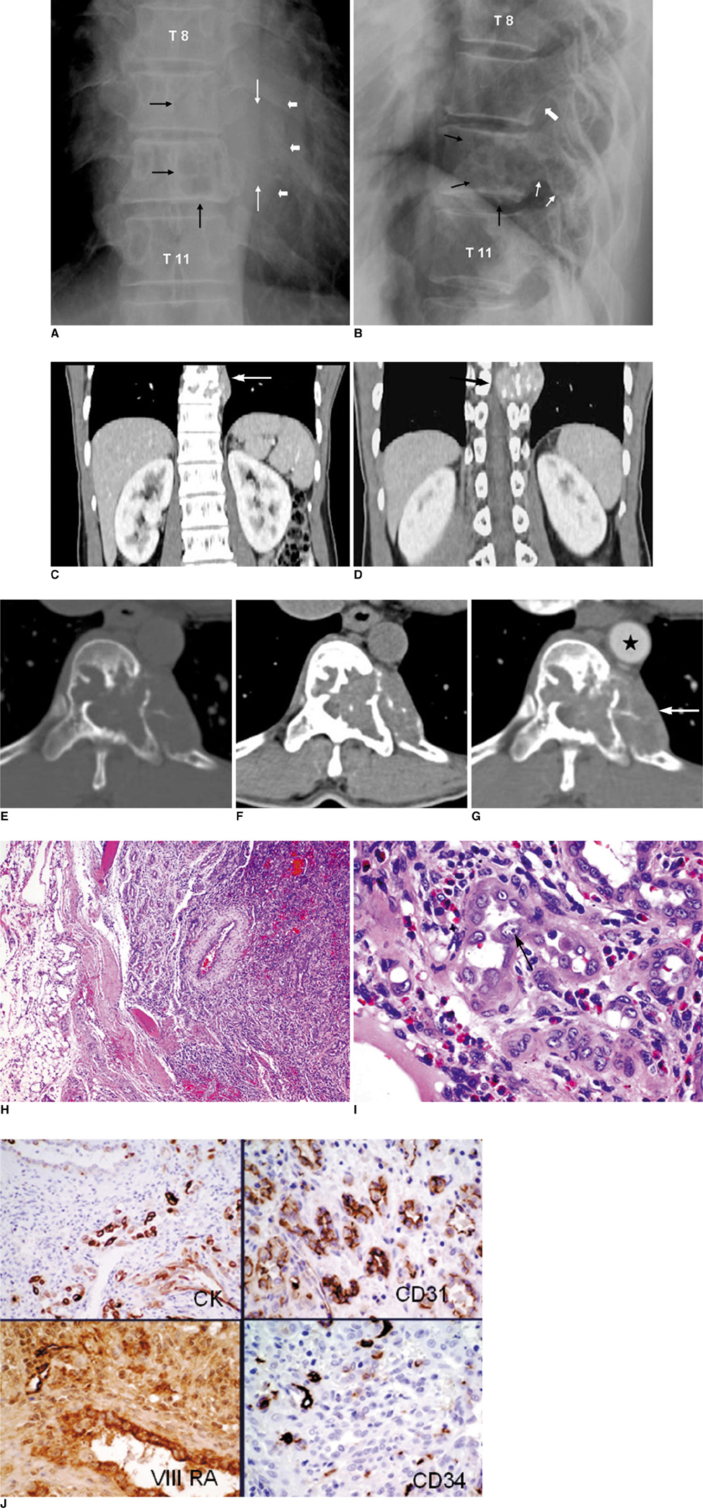
Fig. 1 Epithelioid hemangioma in 48-year-old man. Plain radiographs. Anteroposterior (AP) view (A) reveals geographic osteolytic lesion with partially sclerotic border involving left side of T10 vertebral body. Another similar osteolytic lesion is also observed on left side of T9 vertebral body (black arrows). In addition, permeative osteolytic lesion involving posterior part of left 10th rib at costo-vertebral junction (thin white arrows), associated with soft tissue mass (thick white arrows) was observed. No periosteal reaction is observed. Lateral view (B) reveals lesion involving posterior portion of T10 vertebral body (black arrows) with extension to posterior element (thin white arrows). Osteolytic lesion is also seen involving posterior portion of T9 vertebral body (thick white arrow). C-G. Coronal CT scan (C, D) after iodinated contrast medium injection reveals that lesion involves two contiguous vertebral bodies, T10 and T9 (arrow in C), which causes spinal cord compression (arrow in D). Axial CT scan at T10 vertebral body viewing in bone window (E) and soft tissue window before (F) and after (G) contrast medium injection reveals multiple internal trabeculations, multiple scattered tiny calcifications, and mild enhancement of lesion. Left-side cortex of T10 vertebral body and ventral cortex of 10th rib were destroyed with soft tissue component forming as extrapleural mass (arrow in G) behind thoracic aorta (star). H. Photomicrograph of lesion under low power (×40) reveals well-circumscribed lobular lesion made up of crowded small tubular or gland-like structure surrounded centrally by large vessel with thick wall. Periphery of lesion is surrounded with fibrofatty tissue. I. Under ×400 magnification, lining cells of gland-like structure show irregular vesicular or optically clear nuclei with prominent basophilic nucleoli and occasional vacuolated cytoplasm. Tall and enlarged endothelial cell mimicking 'tomb-stone like structure' projected into lumen, is observed (arrow). J. Immunostains using antibodies reacting to epithelial marker (CK) and vascular markers (CD31, CD34 and Factor VIII RA), show that lining cells are strongly marked by all of these antibodies.
Reference
-
1. Fetsch J. Fletcher C, Unni K, Mertens F, editors. Epitheloid haemangioma. WHO classification of tumors: pathology and genetics of tumors of soft tissue and bones. 2000. Lyon: IARC Press;159–160.2. Sung MS, Kim YS, Resnick D. Epithelioid hemangioma of bone. Skeletal Radiol. 2000. 29:530–534.3. Shanley DJ. Tuberculosis of the spine: imaging features. AJR Am J Roentgenol. 1995. 164:659–664.4. O'Connell JX, Kattapuram SV, Mankin HJ, Bhan AK, Rosenberg AE. Epithelioid hemangioma of bone. A tumor often mistaken for low-grade angiosarcoma or malignant hemangioendothelioma. Am J Surg Pathol. 1993. 17:610–617.5. Ben Romdhane K, Khattech R, Ben Othman M. Epithelioid hemangioma of bone. Am J Surg Pathol. 1994. 18:1270–1271.6. Rosai J, Gold J, Landy R. The histiocytoid hemangiomas. A unifying concept embracing several previously described entities of skin, soft tissue, large vessels, bone, and heart. Hum Pathol. 1979. 10:707–730.7. Ling S, Rafii M, Klein M. Epithelioid hemangioma of bone. Skeletal Radiol. 2001. 30:226–229.8. Jung NY, Jee WH, Ha KY, Park CK, Byun JY. Discrimination of tuberculous spondylitis from pyogenic spondylitis on MRI. AJR Am J Roentgenol. 2004. 182:1405–1410.9. Weiss SW, Goldblum JR. Weiss SW, Goldblum JR, editors. Hemangioendothelioma: vascular tumors of intermediate malignancy. Enzinger and Weiss's soft tissue tumors text book. 2008. Philadelphia: Mosby Elsevier;681–686.10. Weiss SW, Goldblum JR. Weiss SW, Goldblum JR, editors. Epithelioid hemangioma (angiolymphoid hyperplasia with eosinophilia, histiocytoid hemangioma). Enzinger and Weiss's soft tissue tumors text book. 2008. Philadelphia: Mosby Elsevier;644–649.11. Montgomery E. Silverberg SG, DeLellis RA, Frable WJ, editors. Hemangioendothelioma. Silverberg's principles and practice of surgical pathology and cytopathology. 2006. Philadelphia: Churchill Livingstone Elsevier;365–368.12. Nielsen GP, Srivastava A, Kattapuram S, Deshpande V, O'Connell JX, Mangham CD, et al. Epithelioid hemangioma of bone revisited: a study of 50 cases. Am J Surg Pathol. 2009. 33:270–277.
- Full Text Links
-
- Actions
-
Cited
- CITED
-
- Close
- Share
-
- Similar articles
-
- A case of epithelioid sarcoma arising in the vulva
- Multiple Eruptive Epithelioid Hemangiomas on the Right Upper Extremity in an Asian Man
- A Case of Epithelioid Hemangioendothelioma of the External Auditory Canal
- Epithelioid Hemangioendothelioma of the Liver: A Case Report
- Epithelioid Hemangioma of Nasal Tip
