Chest Radiographic Findings in Primary Pulmonary Tuberculosis: Observations from High School Outbreaks
- Affiliations
-
- 1Division of Pulmonary and Critical Care Medicine, Department of Medicine, Samsung Medical Center, Sungkyunkwan University School of Medicine, Seoul 135-710, Korea.
- 2Department of Radiology, Pusan National University Hospital, Pusan National University School of Medicine and Medical Research Institute, Busan 612-617, Korea.
- 3Korean Institute of Tuberculosis, Seoul 121-150, Korea.
- 4Korea Centers for Disease Control and Prevention, Seoul 122-701, Korea.
- 5Department of Radiology, Samsung Medical Center, Sungkyunkwan University School of Medicine, Seoul 135-710, Korea. kyungs.lee@samsung.com
- KMID: 1119223
- DOI: http://doi.org/10.3348/kjr.2010.11.6.612
Abstract
OBJECTIVE
To describe the radiographic findings of primary pulmonary tuberculosis (TB) in previously healthy adolescent patients.
MATERIALS AND METHODS
The Institutional Review Board approved this retrospective study, with a waiver of informed consent from the patients. TB outbreaks occurred in 15 senior high schools and chest radiographs from 58 students with identical strains of TB were analyzed by restriction fragment length polymorphism analysis by two independent observers. Lesions of nodule(s), consolidation, or cavitation in the upper lung zones were classified as typical TB. Mediastinal lymph node enlargement; lesions of nodule(s), consolidation, or cavitation in lower lung zones; or pleural effusion were classified as atypical TB. Inter-observer agreement for the presence of each radiographic finding was examined by kappa statistics.
RESULTS
Of 58 patients, three (5%) had normal chest radiographs. Cavitary lesions were present in 25 (45%) of 55 students. Lesions with upper lung zone predominance were observed in 27 (49%) patients, whereas lower lung zone predominance was noted in 18 (33%) patients. The remaining 10 (18%) patients had lesions in both upper and lower lung zones. Pleural effusion was not observed in any patient, nor was the mediastinal lymph node enlargement. Hilar lymph node enlargement was seen in only one (2%) patient. Overall, 37 (67%) students had the typical form of TB, whereas 18 (33%) had TB lesions of the atypical form.
CONCLUSION
The most common radiographic findings in primary pulmonary TB by recent infection in previously healthy adolescents are upper lung lesions, which were thought to be radiographic findings of reactivation pulmonary TB by remote infection.
MeSH Terms
Figure
-
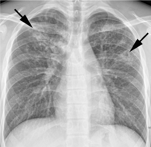
Fig. 1 Primary pulmonary tuberculosis in 18-year-old boy with typical radiographic findings. Chest radiograph shows patchy consolidation, nodules, and cavities (arrows) in bilateral upper lung zones.
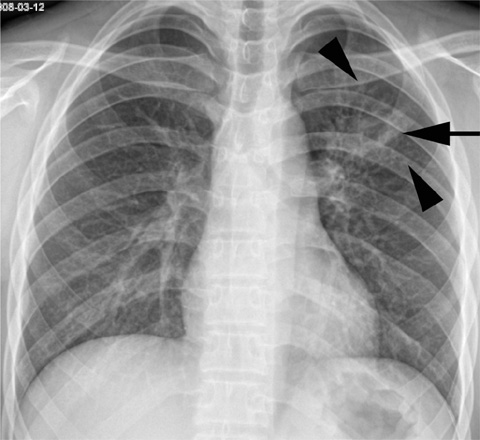
Fig. 2 Pulmonary tuberculosis in 18-year-old boy with typical radiographic findings. Chest radiograph shows cavitary nodule (arrow) with multiple small nodules (arrowheads) in left upper lung zone.
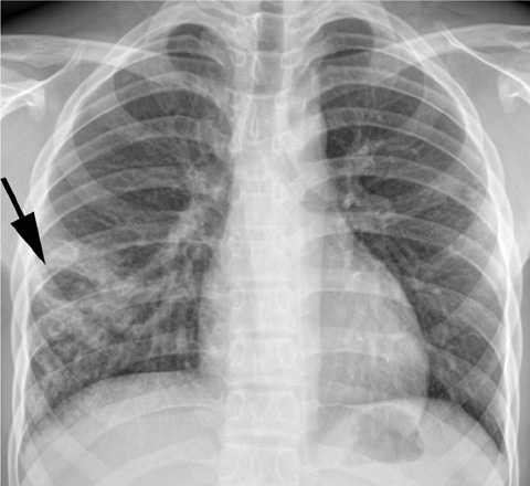
Fig. 3 Pulmonary tuberculosis in 18-year-old boy with atypical radiographic findings. Chest radiograph shows cavitary consolidation (arrow) and nodules in right lower lung zone. Lesions were classified as atypical because they were located in lower lung zone without involvement of upper lung zone.
Cited by 1 articles
-
Clinical Characteristics and Radiologic Patterns of Adelescents with Pulmonary Tuberculosis: Relevance to the Reactive Tuberculosis
Seok Jin Kang, Yeo Hyang Kim, Chi Young Jung, Hee Jung Lee, Myung Chul Hyun
Pediatr Allergy Respir Dis. 2012;22(2):163-170. doi: 10.7581/pard.2012.22.2.163.
Reference
-
1. Diagnostic Standards and Classification of Tuberculosis in Adults and Children. This official statement of the American Thoracic Society and the Centers for Disease Control and Prevention was adopted by the ATS Board of Directors, July 1999. This statement was endorsed by the Council of the Infectious Disease Society of America, September 1999. Am J Respir Crit Care Med. 2000. 161:1376–1395.2. Small PM, Fujiwara PI. Management of tuberculosis in the United States. N Engl J Med. 2001. 345:189–200.3. Lee KS, Song KS, Lim TH, Kim PN, Kim IY, Lee BH. Adult-onset pulmonary tuberculosis: findings on chest radiographs and CT scans. AJR Am J Roentgenol. 1993. 160:753–758.4. Lee JY, Lee KS, Jung KJ, Han J, Kwon OJ, Kim J, et al. Pulmonary tuberculosis: CT and pathologic correlation. J Comput Assist Tomogr. 2000. 24:691–698.5. Jeong YJ, Lee KS. Pulmonary tuberculosis: up-to-date imaging and management. AJR Am J Roentgenol. 2008. 191:834–844.6. Jones BE, Ryu R, Yang Z, Cave MD, Pogoda JM, Otaya M, et al. Chest radiographic findings in patients with tuberculosis with recent or remote infection. Am J Respir Crit Care Med. 1997. 156:1270–1273.7. Geng E, Kreiswirth B, Burzynski J, Schluger NW. Clinical and radiographic correlates of primary and reactivation tuberculosis: a molecular epidemiology study. JAMA. 2005. 293:2740–2745.8. Tabet SR, Goldbaum GM, Hooton TM, Eisenach KD, Cave MD, Nolan CM. Restriction fragment length polymorphism analysis detecting a community-based tuberculosis outbreak among persons infected with human immunodeficiency virus. J Infect Dis. 1994. 169:189–192.9. Small PM, Hopewell PC, Singh SP, Paz A, Parsonnet J, Ruston DC, et al. The epidemiology of tuberculosis in San Francisco. A population-based study using conventional and molecular methods. N Engl J Med. 1994. 330:1703–1709.10. Alland D, Kalkut GE, Moss AR, McAdam RA, Hahn JA, Bosworth W, et al. Transmission of tuberculosis in New York City. An analysis by DNA fingerprinting and conventional epidemiologic methods. N Engl J Med. 1994. 330:1710–1716.11. Hansell DM, Bankier AA, MacMahon H, McLoud TC, Müller NL, Remy J. Fleischner Society: glossary of terms for thoracic imaging. Radiology. 2008. 246:697–722.12. Woodring JH, Vandiviere HM, Fried AM, Dillon ML, Williams TD, Melvin IG. Update: the radiographic features of pulmonary tuberculosis. AJR Am J Roentgenol. 1986. 146:497–506.13. Krysl J, Korzeniewska-Kosela M, Müller NL, FitzGerald JM. Radiologic features of pulmonary tuberculosis: an assessment of 188 cases. Can Assoc Radiol J. 1994. 45:101–107.14. Choyke PL, Sostman HD, Curtis AM, Ravin CE, Chen JT, Godwin JD, et al. Adult-onset pulmonary tuberculosis. Radiology. 1983. 148:357–362.15. Khan MA, Kovnat DM, Bachus B, Whitcomb ME, Brody JS, Snider GL. Clinical and roentgenographic spectrum of pulmonary tuberculosis in the adult. Am J Med. 1977. 62:31–38.16. Kim WS, Choi JI, Cheon JE, Kim IO, Yeon KM, Lee HJ. Primary tuberculosis in infants: radiographic and CT findings. AJR Am J Roentgenol. 2006. 187:1024–1033.17. Leung AN, Müller NL, Pineda PR, FitzGerald JM. Primary tuberculosis in childhood: radiographic manifestations. Radiology. 1992. 182:87–91.18. Sant'Anna C, March MF, Barreto M, Pereira S, Schmidt C. Pulmonary tuberculosis in adolescents: radiographic features. Int J Tuberc Lung Dis. 2009. 13:1566–1568.19. Kim HJ, Lee HJ, Kwon SY, Yoon HI, Chung HS, Lee CT, et al. The prevalence of pulmonary parenchymal tuberculosis in patients with tuberculous pleuritis. Chest. 2006. 129:1253–1258.20. Marais BJ, Parker SK, Verver S, van Rie A, Warren RM. Primary and postprimary or reactivation tuberculosis: time to revise confusing terminology. AJR Am J Roentgenol. 2009. 192:W198.21. Marais BJ, Gie RP, Schaaf HS, Hesseling AC, Obihara CC, Starke JJ, et al. The natural history of childhood intra-thoracic tuberculosis: a critical review of literature from the prechemotherapy era. Int J Tuberc Lung Dis. 2004. 8:392–402.22. Andronikou S, Vanhoenacker FM, De Backer AI. Advances in imaging chest tuberculosis: blurring of differences between children and adults. Clin Chest Med. 2009. 30:717–744.23. Barnes PF, Bloch AB, Davidson PT, Snider DE Jr. Tuberculosis in patients with human immunodeficiency virus infection. N Engl J Med. 1991. 324:1644–1650.24. Leung AN, Brauner MW, Gamsu G, Mlika-Cabanne N, Ben Romdhane H, Carette MF, et al. Pulmonary tuberculosis: comparison of CT findings in HIV-seropositive and HIV-seronegative patients. Radiology. 1996. 198:687–691.25. Newton SM, Brent AJ, Anderson S, Whittaker E, Kampmann B. Paediatric tuberculosis. Lancet Infect Dis. 2008. 8:498–510.
- Full Text Links
-
- Actions
-
Cited
- CITED
-
- Close
- Share
-
- Similar articles
-
- Radiographic Findings of Pulmonary Tuberculosis in Non-AIDS Immunocompromised adult Patients: Comparison with Immunocompetent Adult Patients
- A Study on the Case Rate of Chest Tuberculosis in Female Infertility
- Pulmonary paragonimiasis: CT findings
- Unusual radiological findings of adult-onset pulmonary tuberculosis
- Radiographic Findings of Pulmonary Tuberculosis in Adult Diabetic Patients' Comparison of Diabetics with Nondiabetics of no Other Underlying Diseases
