Fish Bone as a Nidus for Stone Formation in the Common Bile Duct: Report of Two Cases
- Affiliations
-
- 1Department of Diagnostic Radiology, Dongsan Medical Center, Keimyung University College of Medicine, Korea. yhkim68@dsmc.or.kr
- 2Department of Diagnostic Radiology, Andong General Hospital, Korea.
- 3Department of Diagnostic Radiology, Youngnam University Hospital, Korea.
- KMID: 1118826
- DOI: http://doi.org/10.3348/kjr.2004.5.3.210
Abstract
- We report two cases of common bile duct stone formed around a fish bone which migrated from the intestinal tract, along with their characteristic imaging findings. Two patients who had no history of previous operation were admitted because of cholangitis. Percutaneous transhepatic biliary drainage (PTBD) was performed and the cholangiogram showed filling defects with an unusually elongated shape in the common bile duct. After improvement of the cholangitic symptoms, the stones were removed through the PTBD tract under fluoroscopic guidance. A nidus consisting of a 1.5 cm sized fish bone was found in each stone removed.
Keyword
MeSH Terms
Figure
-
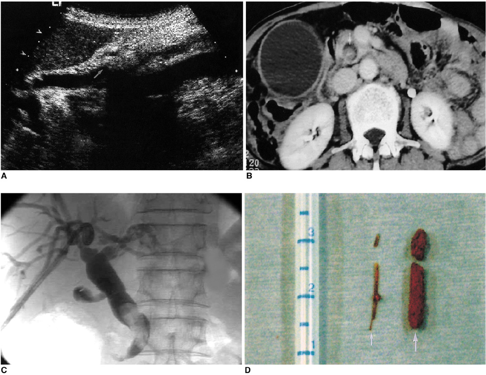
Fig. 1 A 75-year-old woman with common bile duct stone. A. Ultrasonography demonstrates a hyperechoic stone with acoustic shadowing (arrow) in the common bile duct. B. Computed tomography shows a common bile duct stone containing a bright dot of bone density (arrow). C. Cholangiogram shows an elongated filling defect in the common bile duct. D. Photograph of specimen extracted from common bile duct using a stone basket shows a friable pigment stone (thick arrow), that was formed around a fish bone (thin arrow).
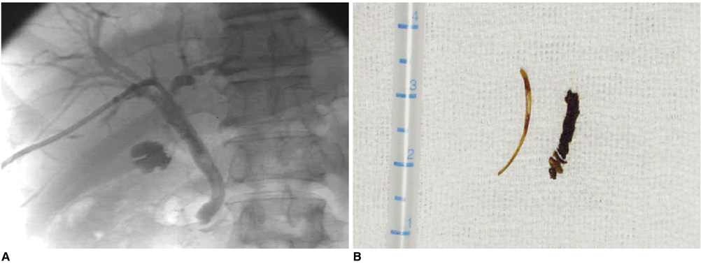
Fig. 2 A 67-year-old man with common bile duct stone. A. Cholangiogram shows an elongated filling defect in the common bile duct. B. Photograph of specimen extracted from common bile duct using a stone basket shows a friable pigment stone, that was formed around a fish bone.
Reference
-
1. Stewart L, Ponce R, Oesterle AL, Griffiss M, Way LW. Pigment gallstone pathogenesis: slime production by biliary bacteria is more important than beta-glucuronidase production. J Gastrointest Surg. 2000. 4:547–553.2. Prochazka V, Krausova D, Kod'ousek R, Zamecnikova P. Foreign material as a cause of choledocholithiasis. Endoscopy. 1999. 31:383–385.3. Orda R, Leviav A, Ratan I, Stadler J, Wiznitzer T. Common bile duct stone caused by foreign body. J Clin Gastroenterol. 1986. 8:466–468.4. McCanse DE, Kurchin A, Hinshaw JR. Gastrointestinal foreign bodies. Am J Surg. 1981. 142:335–337.5. Singh B, Kantu M, Har-El G, Lucente FE. Complications associated with 327 foreign bodies of the pharynx, larynx, and esophagus. Ann Otol Rhinol Laryngol. 1997. 106:301–304.6. Nazoe T, Kitamura M, Adachi Y, et al. Successful conservative treatment for esophageal perforation by a fish bone associated with mediastinitis. Hepatogastroenterology. 1998. 45:2190–2192.7. Solomonov A, Best LA, Goralnik L, Rubin AE, Yigla M. Plerual empyema: an unusual presentation of esophageal perforation. Respiration. 1999. 66:366–368.8. Horii K, Yamazaki O, Matsuyama M, Higaki I, Kawai S, Sakaue Y. Successful treatment of a hepatic abscess that formed secondary to fish bone penetration by percutaneous transhepatic removal of the foreign body: report of a case. Surg Today. 1999. 29:922–926.9. Imamoto T, Tobe T, Mizoguchi K, Ueda T, Igarashi T, Ito H. Perivesical abscess caused by migration of a fish bone from the intestinal tract. Int J Urol. 2002. 9:405–406.10. Lue AJ, Fang WD, Manolidis S. Use of plain radiography and computed tomography to identified fish bone foreign bodies. Otolaryngol Head Neck Surg. 2000. 123:435–438.
- Full Text Links
-
- Actions
-
Cited
- CITED
-
- Close
- Share
-
- Similar articles
-
- Biliary Metal Stent as a Nidus for Bile Duct Stone
- A Common Bile Duct Stone formed by Suture Material after Open Cholecystectomy
- Stent-stone Complex Developed after Long-term Plastic Biliary Stent Placement
- A Case of Common Bile Duct Stone Developed due to a Surgical Clip as a Nidus: An Experience of Successful Management by Endoscopy
- A Comparative Study of Bile Compositions From patients with Gallbladder Stones and Common Bile Duct stones
