The Value of Ultrasound-Guided Tattooing Localization of Nonpalpable Breast Lesions
- Affiliations
-
- 1Department of Radiology and Center for Breast Cancer, National Cancer Center, Goyang-si, Korea. bkhan@smc.samsung.co.kr
- 2Department of Radiology and Center for Imaging Science, Samsung Medical Center, Sungkyunkwan University School of Medicine, Seoul, Korea.
- 3Department of General Surgery, Samsung Medical Center, Sungkyunkwan University School of Medicine, Seoul, Korea.
- KMID: 1110726
- DOI: http://doi.org/10.3348/kjr.2007.8.4.295
Abstract
OBJECTIVE
To investigate the value of ultrasound-guided tattooing localization (US-tattoo) using a charcoal suspension for breast lesions. MATERIALS AND METHODS: One hundred sixty-four nonpalpable breast lesions in 134 patients (mean age 47 years; range 30-74 years) were marked with a charcoal suspension under US guidance. The medical records associated with the US-tattoo, the pathology results and the follow-up US results were reviewed. RESULTS: The average size of the localized lesions was 1.0 cm. The procedure time was < 5 minutes (range, 2-10 minutes) per lesion. The US-tattoo was well tolerated in all cases. The only technical difficulty encountered was a needle tip blockage caused by a large charcoal particle (4.9%). The surgeon easily identified the tattoo with the exception one case. In addition, surgery could be safely delayed from one to 57 days after the making US-tattoo. The pathology result was benign in 108 cases, borderline in five, and malignant in 51. The excised specimen was < 4 cm in 76.6% (82/107) of the benign cases (mean; 2.7 cm). The pathologist could identify the mass around the tattoo and was able to make a specific diagnosis in 81.3% (87/107) of benign lesions. The only complication encountered was residual charcoal marking along the incision scar (3.6%). All follow-up US documented the removal of the lesions. CONCLUSION: An US-tattoo for nonpalpable breast lesions is a very simple and accurate method that can help surgeons design and schedule an open biopsy.
MeSH Terms
Figure
-
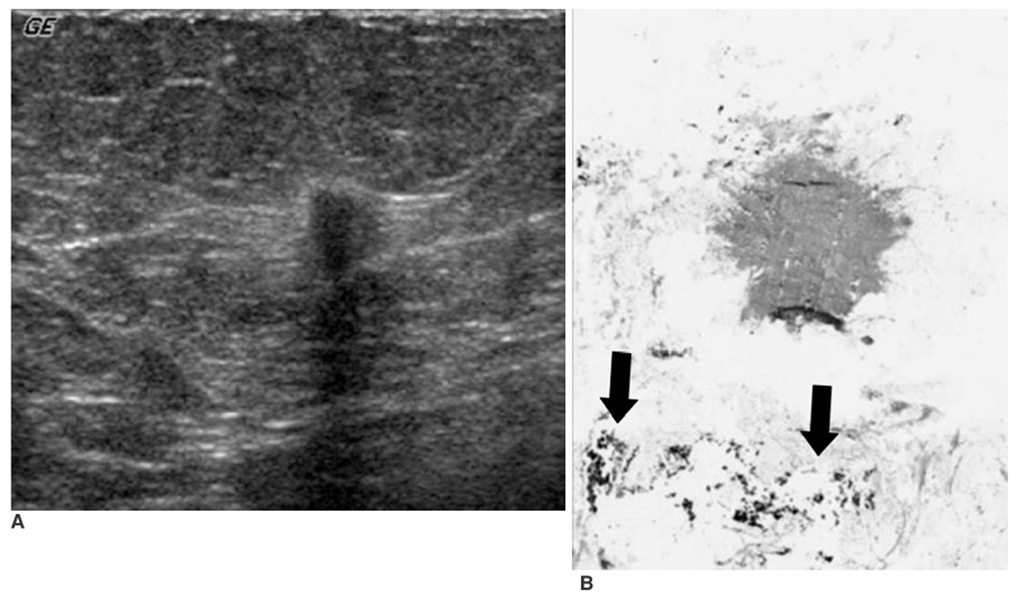
Fig. 1 A 67-year-old woman with a dense pattern on the screening mammogram. A. Ultrasonogram shows a 0.5 cm-sized, irregular taller shaped and hypoechoic solid nodule. B. Photomicrograph of the specimen after the US-tattoo shows a small infiltrative ductal cancer. Adjacent to the mass, charcoal markings are observed as black particles (black arrows). (Hematoxylin & Eosin staining; original magnification, ×40)
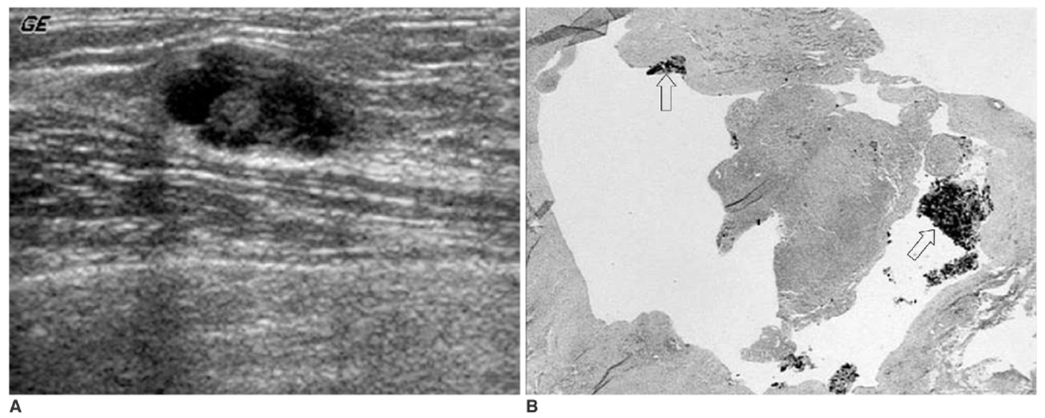
Fig. 2 A 31-year-old woman presented for screening. A. Ultrasonogram shows a 1.3-cm-sized, complex cystic mass. B. Photomicrograph of the specimen after the US-tattoo shows a cystically dilated duct, containing charcoal (white arrows) and inflammatory exudates. (Hematoxylin & Eosin staining; original magnification, ×40)
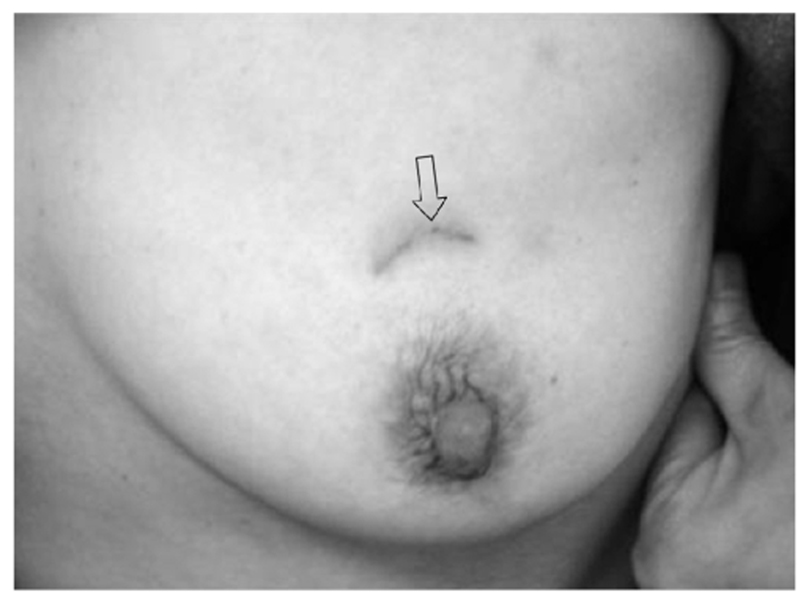
Fig. 3 Photograph of the left breast after breast conserving surgery demonstrates a residual charcoal stain along the scar.
Reference
-
1. Altomare V, Guerriero G, Giacomelli L, Battista C, Carino R, Montesano M, et al. Management of nonpalpable breast lesions in a modern functional breast unit. Breast Cancer Res Treat. 2005. 93:85–89.2. Czarnecki DJ, Feider HK, Splittgerber GF. Toluidine blue dye as a breast localization marker. AJR Am J Roentgenol. 1989. 153:261–263.3. Homer MJ, Smith TJ, Safaii H. Prebiopsy needle localization. Methods, problems, and expected results. Radiol Clin North Am. 1992. 30:139–153.4. Rosenberg AL, Schwartz GF, Feig SA, Patchefsky AS. Clinically occult breast lesions: localization and significance. Radiology. 1987. 162:167–170.5. Saarela AO, Kiviniemi HO, Rissanen TJ. Preoperative methylene blue staining of galactographically suspicious breast lesions. Int Surg. 1997. 82:403–405.6. Svane G. A stereotaxic technique for preoperative marking of non-palpable breast lesions. Acta Radiol Diagn (Stockh). 1983. 24:145–151.7. Bristol JB, Jones PA. Transgression of localizing wire into the pleural cavity prior to mammography. Br J Radiol. 1981. 54:139–140.8. Davis PS, Wechsler RJ, Feig SA, March DE. Migration of breast biopsy localization wire. AJR Am J Roentgenol. 1988. 150:787–788.9. Helvie MA, Ikeda DM, Adler DD. Localization and needle aspiration of breast lesions: complications in 370 cases. AJR Am J Roentgenol. 1991. 157:711–714.10. Homer MJ. Transection of the localization hooked wire during breast biopsy. AJR Am J Roentgenol. 1983. 141:929–930.11. Canavese G, Catturich A, Vecchio C, Tomei D, Estienne M, Moresco L, et al. Pre-operative localization of non-palpable lesions in breast cancer by charcoal suspension. Eur J Surg Oncol. 1995. 21:47–49.12. Langlois SL, Carter ML. Carbon localisation of impalpable mammographic abnormalities. Australas Radiol. 1991. 35:237–241.13. Sperry K. Tattoos and tattooing. Part I: History and methodology. Am J Forensic Med Pathol. 1991. 12:313–319.14. Bonhomme-Faivre L, Mathieu MC, Grossiord JL, Depreatere P, Couarraze G, Orbach-Arbouys S, et al. Formulation of a charcoal suspension for intratumor injection. Part 1: Study of the nature, granulometry, and concentration. Pharm Res. 1997. 14:218–223.15. Bonhomme-Faivre L, Depraetere P, Savelli MP, Amdidouche D, Bizi E, Seiller M, et al. Charcoal suspension for tumor labelling modifies macrophage activity in mice. Life Sci. 2000. 66:817–827.16. Mullen DJ, Eisen RN, Newman RD, Perrone PM, Wilsey JC. The use of carbon marking after stereotactic large-core-needle breast biopsy. Radiology. 2001. 218:255–260.17. Burbank F, Forcier N. Tissue marking clip for stereotactic breast biopsy: initial placement accuracy, long-term stability, and usefulness as a guide for wire localization. Radiology. 1997. 205:407–415.18. Fajardo LL, Bird RE, Herman CR, DeAngelis GA. Placement of endovascular embolization microcoils to localize the site of breast lesions removed at stereotactic core biopsy. Radiology. 1998. 206:275–278.19. Kopans DB. Review of stereotaxic large-core needle biopsy and surgical biopsy results in nonpalpable breast lesions. Radiology. 1993. 189:665–666.20. Liberman L, Dershaw DD, Morris EA, Abramson AF, Thornton CM, Rosen PP. Clip placement after stereotactic vacuum-assisted breast biopsy. Radiology. 1997. 205:417–422.
- Full Text Links
-
- Actions
-
Cited
- CITED
-
- Close
- Share
-
- Similar articles
-
- Usefulness of Ultrasound-Guided Automated Core Biopsy of Nonpalpable Breast Lesions
- The Results of Mammography and Ultrasound-Guided Localization Biopsy of Nonpalpable Breast Lesions, and the Differences between Them
- Values of US-Guided Localization and Specimen US in Nonpalpable Breast Masses
- Effectiveness of preoperative ultrasound-guided charcoal tattooing for localization of metastatic melanoma
- US-Guided Preoperative Hook-Wire Localization of Nonpalpable Breast Lesions
