Time of initial detection of fetal and extra-fetal structures by ultrasonographic examination in Miniature Schnauzer bitches
- Affiliations
-
- 1College of Veterinary Medicine, Chonnam National University, Gwangju 500-757, Korea. chson@chonnam.ac.kr
- KMID: 1090805
- DOI: http://doi.org/10.4142/jvs.2007.8.3.289
Abstract
- Serial ultrasonographic examinations were performeddaily on 9 Miniature Schnauzer bitches from the 15th dayof gestation until parturition to determine the time thegestational structures were first detected. The gestationalage was timed from the day of ovulation (day 0), whichwas estimated to occur when the plasma progesteroneconcentration was >4.0ng/ml. The gestational length in 9Miniature Schnauzer bitches was found to be 63.0+/-1.7(range 61-65) days. The initial detection of the fetal andextra-fetal structures were as follows: gestational sac atday 18.0+/-0.9 (17-19); zonary placenta in the uterine wallat day 24.9+/-1.1 (23-26); yolk sac membrane at day 25.0+/-0.9 (24-26); amnionic membrane at day 27.7+/-1.0 (26-29); embryo initial detection at day 22.6+/-0.5 (22-23);heartbeat at day 23.4+/-0.5 (23-24); fetal movement at day32.5+/-0.8 (32-34); stomach at day 31.2+/-1.6 (29-33);urinary bladder at day 32.6+/-1.8 (31-35); skeleton at day34.9+/-1.6 (34-38) and kidney at day 42.2+/-0.7 (41-43).
MeSH Terms
Figure
-
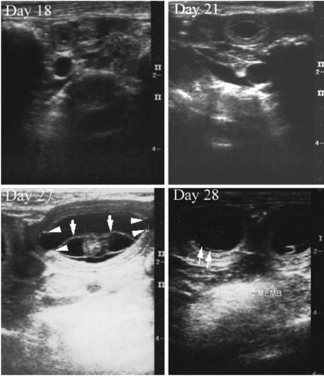
Fig. 1 Ultrasonograms of the extra-fetal structures in pregnant Miniature Schnauzer bitches. Day 18: Transverse image of the first detection of an anechoic gestational sac. Day 21: Longitudinal image of the gestational sac. An echogenic inner placental layer was detected in the uterine wall. Day 27: Longitudinal image of the gestational sac contained an embryo and the tubular shape of the yolk sac membrane (white arrows). The zonary placenta (white arrowheads) was cylindrical in shape and appeared folded inward at the edges. Day 28: The amnionic membrane (white arrows) appeared faint.
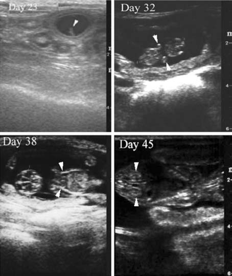
Fig. 2 Ultrasonograms of the fetal structures in pregnant Miniature Schnauzer bitches. Day 23: Transverse image of the gestational sac contained an oblong-shaped embryo (white arrowhead) apposed to the uterine wall. Day 32: Longitudinal image of a fetus with an anechoic area in the head and forelimb buds (white arrowheads). The fetal crown-rump length was 20 mm. Day 38: Longitudinal image of a fetus with hyperechoic skeletal structures in the head and thoracic wall (white arrowheads). Day 45: Longitudinal image of a fetal vertebral column and kidney (white arrowheads).
Reference
-
1. Boyd JS, Renton JP, Harvey MJ, Nickson DA, Eckersall PD, Ferguson JM. Problems associated with ultrasonography of the canine ovary around the time of ovulation. J Reprod Fertil Suppl. 1993. 47:101–105.2. Christiansen IJ. Reproduction in the Dog and Cat. 1984. 1st ed. London: Baillière Tindall;118–119.3. Concannon P, Rendano V. Radiographic diagnosis of canine pregnancy: onset of fetal skeletal radiopacity in relation to times of bleeding, preovulatory luteinizing hormone release, and parturition. Am J Vet Res. 1983. 44:1506–1511.4. England GCW, Allen EW. Studies on canine pregnancy using B-mode ultrasound: Diagnosis of early pregnancy and the number of conceptuses. J Small Anim Pract. 1990. 31:321–323.
Article5. England GCW, Porter DJ. Studies on canine pregnancy using B-mode ultrasound: development of the conceptus and determination of gestational age. J Small Anim Pract. 1990. 31:324–329.
Article6. England GCW, Russo M. Ultrasonographic characteristics of early pregnancy failure in bitches. Theriogenology. 2006. 66:1694–1698.
Article7. England GCW, Yeager AE. Ultrasonographic appearance of the ovary and uterus of the bitch during oestrus, ovulation and early pregnancy. J Reprod Fertil Suppl. 1993. 47:107–117.8. Holst PA, Phemister RD. Onset of diestrus in the Beagle bitch: definition and significance. Am J Vet Res. 1974. 35:401–406.9. Kang BK, Choi HS, Son CH, Shin CR, Seo DH, Park IC. Ultrasonographic apperance of the gestational structures throughout pregnancy in pet dogs. I. Time of initial detection of the fetal and extra-fetal structures. Korean J Vet Clin Med. 1997. 14:279–286.10. Kim JH, Jeong KA, Kang HG, Oh KS, Park IC, Park SG, Han HJ, Son CH. Relationship between vaginal cytology and reproductive hormone during the estrous cycle in Korea Jin-do bitches. Korean J Vet Clin Med. 2000. 17:225–233.11. Ko JS, Kim BS, Lee SA, Cho YT, Kim JP, Oh KS, Kim SH, Kim JT, Park IC, Kim YH, Son CH. Ultrasonographic apperance of the gestational structures throughout pregnancy in Shih-tzu bitches. I. Time of initial detection of the fetal and extra-fetal structures. Korean J Vet Clin. 2004. 21:29–34.12. Luvoni GC, Beccaglia M. The prediction of parturition date in canine pregnancy. Reprod Domest Anim. 2006. 41:27–32.
Article13. Luvoni GC, Grioni A. Determination of gestational age in medium and small size bitches using ultrasonographic fetal measurements. J Small Anim Pract. 2000. 41:292–294.
Article14. Moriyoshi M, Waki Y, Nakao T, Kawata K. Observation of the growth process of a beagle embryo and fetus by ultrasonography. J Vet Med Sci. 1996. 58:443–445.
Article15. Okkens AC, Teunissen JM, Van Osch W, Van Den Brom WE, Dieleman SJ, Kooistra HS. Influence of litter size and breed on the duration of gestation in dogs. J Reprod Fertil Suppl. 2001. 57:193–197.16. Pharr JW, Post K. Ultrasonography and radiography of the canine postpartum uterus. Vet Radiol Ultrasound. 1992. 33:35–40.
Article17. Schutte AP. Canine vaginal cytology. I. Technique and cytological morphology. J Small Anim Pract. 1967. 8:301–306.18. Son CH, Jeong KA, Kim JH, Park IC, Kim SH, Lee CS. Establishment of the prediction table of parturition day with ultrasonography in small pet dogs. J Vet Med Sci. 2001. 63:715–721.
Article19. Wallace SS, Mahaffey MB, Miller DM, Thompson FN, Chakraborty PK. Ultrasonographic appearance of the ovaries of dogs during the follicular and luteal phases of the estrous cycle. Am J Vet Res. 1992. 53:209–215.20. Yeager AE, Mohammed HO, Meyers-Wallen V, Vannerson L, Concannon PW. Ultrasonographic appearance of the uterus, placenta, fetus, and fetal membranes throughout accurately timed pregnancy in beagels. Am J Vet Res. 1992. 53:342–351.
- Full Text Links
-
- Actions
-
Cited
- CITED
-
- Close
- Share
-
- Similar articles
-
- A case of fetal intracranial hemorrhage diagnosed by antenatal ultrasonographic examination
- Fetal Fibronectin and Ultrasonographic Examination of Cervix: Which is Better as a Predictor of Preterm Delivery in Patients with Preterm Labor and Intact Membranes?
- The determination of dark adaptation time using electroretinography in conscious Miniature Schnauzer dogs
- The availability of ultrasonography as the method for early detection of fetal chromosomal abnormalities
- Technique of fetal echocardiography
