Primary Extraskeletal Mesenchymal Chondrosarcoma Arising from the Pancreas
- Affiliations
-
- 1Department of Radiology, Ilsan Paik Hospital, Inje University, School of Medicine, Goyang-si, Gyeonggi-do , Korea. hanyoonhee@ilsanpaik.ac.kr
- 2Department of Pathology, Ilsan Paik Hospital, Inje University, School of Medicine, Goyang-si, Gyeonggi-do, Korea.
- KMID: 1089442
- DOI: http://doi.org/10.3348/kjr.2007.8.6.541
Abstract
- We report here on a case of primary extraskeletal mesenchymal chondrosarcoma that arose from the pancreas. A 41-year-old man was evaluated by CT to find the cause of his abdominal pain. The CT scans showed a heterogeneously enhancing necrotic mass with numerous areas of coarse calcification, and this was located in the left side of the retroperitoneal space and involved the body and tail of the pancreas. Portal venography via the celiac axis also showed invasion of the splenic vein. Following excision of the mass, it was pathologically confirmed to be primary extraskeletal mesenchymal chondrosarcoma that arose from the pancreas.
Keyword
MeSH Terms
-
Abdominal Pain/etiology
Adult
Chondrosarcoma, Mesenchymal/complications/*diagnosis/surgery
Contrast Media/administration & dosage
Diagnosis, Differential
Humans
Iohexol/analogs & derivatives/diagnostic use
Male
Necrosis
Pancreas/pathology/radiography
Pancreatic Neoplasms/complications/*diagnosis/surgery
Portal Vein/radiography
Radiographic Image Enhancement/methods
Rare Diseases
Retroperitoneal Space/radiography
Splenic Vein/radiography
Tomography, X-Ray Computed/methods
Figure
-
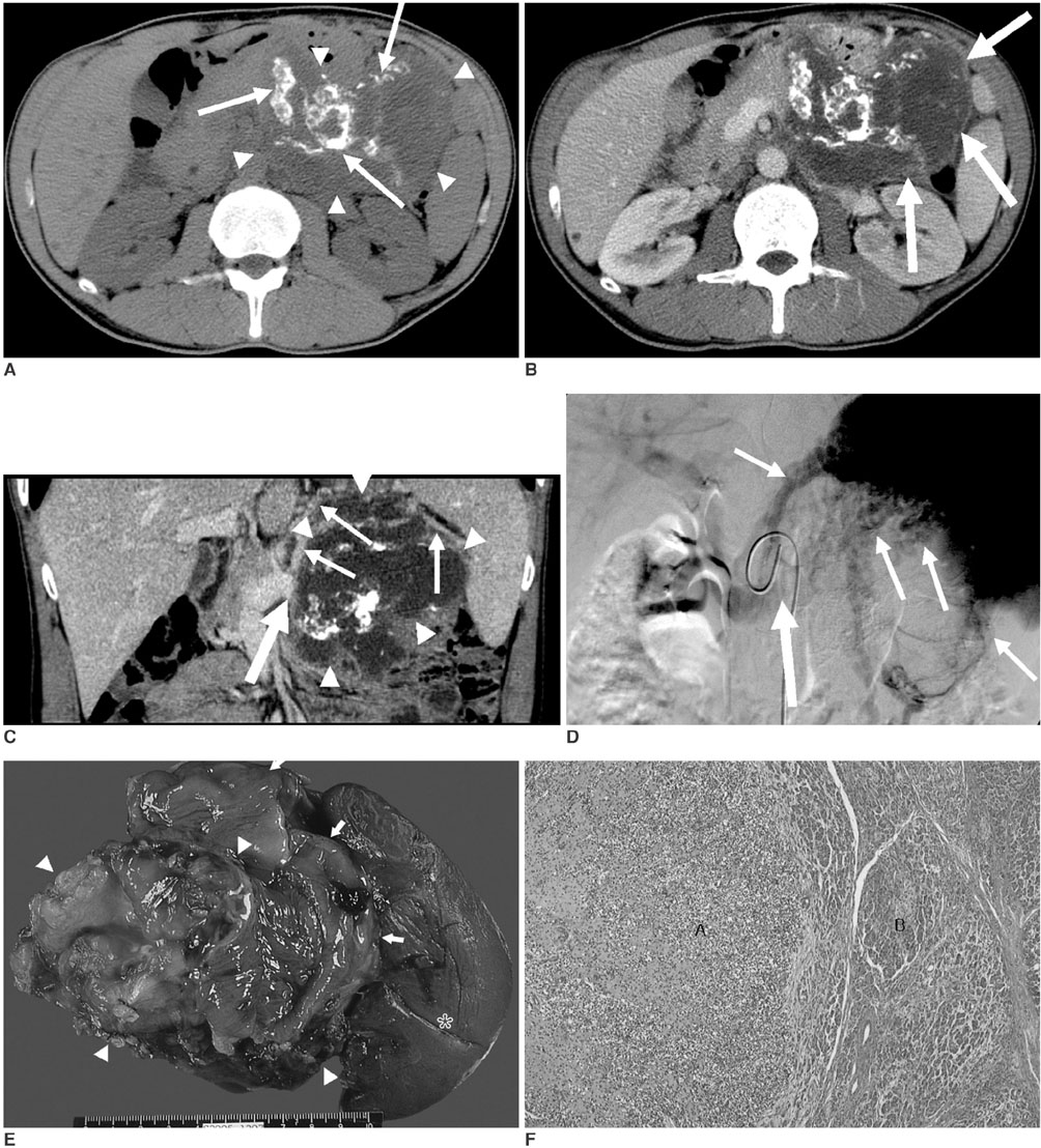
Fig. 1 A. Unenhanced transverse CT scan reveals an ill defined, lobulated mass (arrowheads) of heterogeneously low attenuation that contains numerous areas of coarse calcification (arrows) in the left side of the retroperitoneum. B. On the contrast-enhanced transverse CT scan obtained during the portal venous phase, subtle enhancement is only noted in the periphery of the mass (arrows), and most of the mass remains without enhancement. C. The contrast-enhanced coronal reformatted image shows the ill defined necrotic low attenuated mass (arrowheads) and obliteration of the splenic vein by the mass (thick arrow) with the development of multiple collateral veins (thin arrows). D. Portal venography via the celiac axis also shows obliteration of the splenic vein (thick arrow) by the mass and the development of multiple collateral veins (thin arrows). E. A large multilobulated mass (arrowheads) is located in the body and tail of the pancreas with attachment to the spleen (asterisk) and transverse colon (arrows). F. Light microscopic image shows the typical bimorphic pattern of chondrosarcoma. The chondroid zone (A) is surrounded by a proliferation of undifferentiated cells (B) with abrupt transition. (Hematoxylin & Eosin staining, ×100)
Reference
-
1. González-Cámpora R, Otal Salaverri C, Gomez Pascual A, Hevia Vazquez A, Galera Davidson H. Mesenchymal chondrosarcoma of the retroperitoneum. Report of a case diagnosed by fine needle aspiration biopsy with immunohistochemical, electron microscopic demonstration of S-100 protein in undifferentiated cells. Acta Cytol. 1995. 39:1237–1243.2. Shapeero LG, Vanel D, Couanet D, Contesso G, Ackerman LV. Extraskeletal mesenchymal chondrosarcoma. Radiology. 1993. 186:819–826.3. Komatsu T, Taira S, Matsui O, Takashima T, Note M, Fujita H. A case of ruptured mesenchymal chondrosarcoma of the pancreas. Radiat Med. 1999. 17:239–241.4. Mikhail MG, Lim KB. Dedifferentiated chondrosarcoma metastasizing to the pancreas in pregnancy. Acta Obstet Gynecol Scand. 1989. 68:467–468.5. Lichtenstein L, Bernstein D. Unusual benign and malignant chondroid tumors of bone. A survey of some mesenchymal cartilage tumors and malignant chondroblastic tumors, including a few multicentric ones, as well as many atypical benign chondroblastomas and chondromyxoid fibromas. Cancer. 1959. 12:1142–1157.6. Dowling EA. Mesenchymal chondrosarcoma. J Bone Joint Surg Am. 1964. 46:747–754.7. Doria MI Jr, Wang HH, Chinoy MJ. Retroperitoneal mesenchymal chondrosarcoma. Report of a case diagnosed by fine needle aspiration cytology. Acta Cytol. 1990. 34:529–532.8. Louvet C, de Gramont A, Krulik M, Jagueux M, Hubert D, Brissaud P, et al. Extraskeletal mesenchymal chondrosarcoma: case report and review of the literature. J Clin Oncol. 1985. 3:858–863.9. White DW, Ly JQ, Beall DP, McMillan MD, McDermott JH. Extraskeletal mesenchymal chondrosarcoma: case report. Clin Imaging. 2003. 27:187–190.10. Nakashima Y, Unni KK, Shives TC, Swee RG, Dahlin DC. Mesenchymal chondrosarcoma of bone and soft tissue. A review of 111 cases. Cancer. 1986. 57:2444–2453.
- Full Text Links
-
- Actions
-
Cited
- CITED
-
- Close
- Share
-
- Similar articles
-
- A Case of Mesenchymal Chondrosarcoma In Pancreas
- Extraskeletal Mesenchymal Chondrosarcoma of the Mediastinum: A Case Report
- A Case of Postoperative Chemotherapy of Extraskeletal Mesenchymal Chondrosarcoma
- Extraskeletal Mesenchymal Chondrosarcoma of the Carotid Space: A Case Report
- Primary extraskeletal mesenchymal chondrosarcoma of the vulva
