Anat Cell Biol.
2017 Jun;50(2):86-92. 10.5115/acb.2017.50.2.86.
Study of sexual dimorphism of Malaysian crania: an important step in identification of the skeletal remains
- Affiliations
-
- 1Forensic Unit, Department of Pathology, Universiti Kebangsaan Malaysia Medical Centre, Kuala Lumpur, Malaysia. faridah.nor@ukm.edu.my
- 2Department of Forensic Medicine and Clinical Toxicology, Faculty of Medicine, Al Azhar University, Cairo, Egypt.
- 3Faculty of Dentistry, Universiti Sains Islam Malaysia, Kuala Lumpur, Malaysia.
- 4Department of Anatomy, Universiti Kebangsaan Malaysia Medical Centre, Kuala Lumpur, Malaysia.
- KMID: 2451232
- DOI: http://doi.org/10.5115/acb.2017.50.2.86
Abstract
- Sex determination is one of the main steps in the identification of human skeletal remains. It constitutes an initial step in personal identification from the skeletal remains. The aim of the present study was to provide the population-specific sex discriminating osteometric standards to aid human identification. The present study was conducted on 87 (174 sides) slices of crania using postmortem computed tomography in 45 males and 42 females, aged between 18 and 75 years. About 22 parameters of crania were measured using Osirix software 3-D Volume Rendering. Results showed that all parameters were significantly higher in males than in females except for orbital height of the left eye by independent t test (P<0.01). By discriminant analysis, the classification accuracy was 85.1%, and by regression, the classification accuracy ranged from 78.2% to 86.2%. In conclusion, cranium can be used to distinguish between males and females in the Malaysian population. The results of the present study can be used as a forensic tool for identification of unknown crania.
Keyword
Figure
-
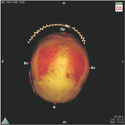
Fig. 1 Superior view of the crania. AS, anteriorly superior; Eu, euryon; G, glabella; L, lateral; Op, opisthocranion; PI, posteriorly inferior; R, right.
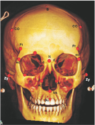
Fig. 2 Frontal view of the crania. AI, alare; B, bregma; CO, coronalia; d, dacryon; ec, ectoconchion; Ft, frontotempolare; N, nasion; Ns, nasospinale; Pr, prosthions; Zy, zygomatic arch.
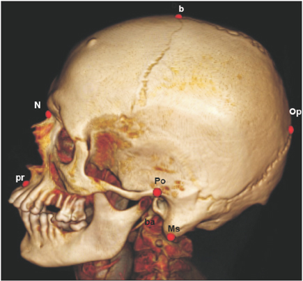
Fig. 3 Lateral view of the crania. b, bregma; ba, basion; Ms, mastoidale; N, nasion; Op, opithocranion; Po, porion; Pr, prosthion.
Reference
-
1. Krogman W, Īşcan MY. The human skeleton in forensic medicine. 2nd ed. Springfield, IL: Charles C. Thomas;1986.2. Scheuer L. Application of osteology to forensic medicine. Clin Anat. 2002; 15:297–312.3. Loth SR, Iscan MY. Sex determination. In : Siegal AJ, Saukko PJ, Knupfer GC, editors. Encyclopedia of Forensic Sciences. San Diego, CA: Academic Press;2000. p. 252–260.4. Patil KR, Mody RN. Determination of sex by discriminant function analysis and stature by regression analysis: a lateral cephalometric study. Forensic Sci Int. 2005; 147:175–180.5. Suazo GI, Zavando MD, Smith RL. Performance evaluation as a diagnostic test for traditional methods for forensic identification of sex. Int J Morphol. 2009; 27:381–386.6. Suazo GI, Zavando D. Age effect in the morphological traits performance for sex determination in human skulls and mandibles. Int J Morphol. 2012; 30:296–301.7. Spradley MK, Jantz RL. Sex estimation in forensic anthropology: skull versus postcranial elements. J Forensic Sci. 2011; 56:289–296.8. Iscan MY, Steyn M. The human skeleton in forensic medicine. Springfield, IL: Charles C. Thomas;2013.9. Franklin D, Freedman L, Milne N. Three-dimensional technology for linear morphological studies: a re-examination of cranial variation in four southern African indigenous populations. Homo. 2005; 56:17–34.10. Ekizoglu O, Hocaoglu E, Inci E, Can IO, Solmaz D, Aksoy S, Buran CF, Sayin I. Assessment of sex in a modern Turkish population using cranial anthropometric parameters. Leg Med (Tokyo). 2016; 21:45–52.11. IBM Corp. Released 2013. IBM SPSS Statistics for Windows, version 23.0. Armonk, NY: IBM Corp;2015.12. Brown MT, Wicker LR. Discriminant analysis. In : Tinsley HE, Brown SD, editors. Handbook of Applied Multivariate Statistics and Mathematical Modeling. San Diego, CA: Academic Press;2000. p. 209–235.13. Gapert R, Black S, Last J. Sex determination from the foramen magnum: discriminant function analysis in an eighteenth and nineteenth century British sample. Int J Legal Med. 2009; 123:25–33.14. Franklin D, Freedman L, Milne N. Sexual dimorphism and discriminant function sexing in indigenous South African crania. Homo. 2005; 55:213–228.15. Franklin D, Freedman L, Milne N, Oxnard CE. A geometric morphometric study of sexual dimorphism in the crania of indigenous southern Africans. S Afr J Sci. 2006; 102:229–238.16. Dayal MR, Spocter MA, Bidmos MA. An assessment of sex using the skull of black South Africans by discriminant function analysis. Homo. 2008; 59:209–221.17. Kranioti EF, Işcan MY, Michalodimitrakis M. Craniometric analysis of the modern Cretan population. Forensic Sci Int. 2008; 180:110.e1–110.e5.18. Hanihara K. Sex diagnosis of Japanese skulls and scapulae. J Anthropol Soc Nippon. 1959; 67:191–197.19. Giles E, Elliot O. Sex determination by discriminant function analysis of crania. Am J Phys Anthropol. 1963; 21:53–68.20. Chovalopoulou ME, Valakos ED, Manolis SK. Sex determination by three-dimensional geometric morphometrics of the palate and cranial base. Anthropol Anz. 2013; 70:407–425.21. Iscan MY, Steyn M. Craniometric determination of population affinity in South Africans. Int J Legal Med. 1999; 112:91–97.22. Steyn M, Işcan MY. Sexual dimorphism in the crania and mandibles of South African whites. Forensic Sci Int. 1998; 98:9–16.23. Ogawa Y, Imaizumi K, Miyasaka S, Yoshino M. Discriminant functions for sex estimation of modern Japanese skulls. J Forensic Leg Med. 2013; 20:234–238.
- Full Text Links
-
- Actions
-
Cited
- CITED
-
- Close
- Share
-
- Similar articles
-
- Study of sexual dimorphism of Malaysian crania: an important step in identification of the skeletal remains
- Erratum: Study of sexual dimorphism of Malaysian crania: an important step in identification of the skeletal remains
- Craniometric study for sex determination in a Thai population
- Sex estimation from upper limb bones in a Thai population
- Sex estimation using subpubic angle from reconstructed three-dimensional computed tomography pelvic model in a contemporary Malaysian population
