Evolution of the paranasal sinuses' anatomy through the ages
- Affiliations
-
- 1Department of Anatomy, Medical Faculty, Aristotle University of Thessaloniki, Thessaloniki, Greece. g_paraskevas@yahoo.gr
- KMID: 2046763
- DOI: http://doi.org/10.5115/acb.2013.46.4.235
Abstract
- Previously, anatomists considered paranasal sinuses as a mysterious region of the human skull. Historically, paranasal sinuses were first identified by ancient Egyptians and later, by Greek physicians. After a long period of no remarkable improvement in the understanding of anatomy during the Middle Ages, anatomists of the Renaissance period-Leonardo da Vinci and Vesalius-made their own contribution. Nathaniel Highmore's name is also associated with the anatomy of paranasal sinuses as he was first to describe the maxillary sinus.
Keyword
Figure
-

Fig. 1 Leonardo da Vinci's depiction of a skull. The image is one of da Vinci's anatomical drawings of a human skull. The left half of the skull is sectioned to reveal the frontal sinus and the maxillary sinus. Of note is the close relation of the two sinuses to the orbit and the teeth of the upper jaw, as understood by Leonardo. Reprinted from Leonardo Da Vinci's drawings [11].
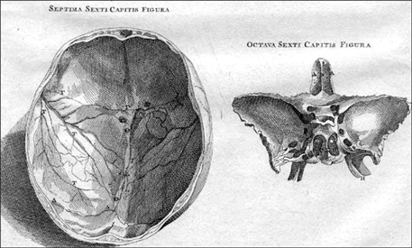
Fig. 2 Vesalius' images of the skull and the sphenoid bone. The images constitute anatomical drawings from Vesalius' work 'De Humani Corporis Fabrica.' Most of the illustrations in this book were created by Jan Stephan van Calcar, an Italian artist, one of Titian's students. The left half of the picture shows a transverse cross-section of the skull, depicting the calvaria whereas the right half shows the sphenoid bone. The right-hand image shows the frontal sinus as well as the two sphenoid sinuses, which are separated by the sphenoid septum. Reprinted from Andreas Vesalius, De Humani Corporis Fabrica [13].
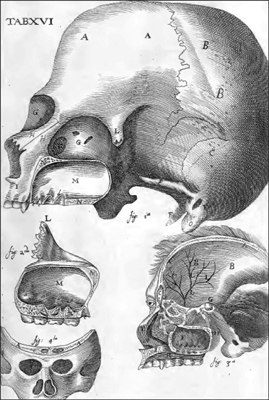
Fig. 3 Illustrations from Nathaniel Highmore's book 'Corporis Humani Disquisitio Anatomica.' The maxillary sinus and the projection of the teeth of the upper jaw into the floor of the sinus are clearly shown. The cross-section of the skull in the bottom right of the picture reveals the frontal sinus, sphenoid sinus, ethmoid cells and maxillary sinus. Reprinted from Nathaniel Highmore, Corporis humani disquisitio anatomica, p. 227 [19].
Reference
-
1. Nall GH. Macmillan's elementary Latin-English dictionary. London: Macmillan and Co. Ltd;1964.2. Gazi A. Dictionary of the Greek language. Athens: Mati;1839.3. Formby ML. The maxillary sinus. Proc R Soc Med. 1960; 53:163–168.4. Lascaratos JG, Segas JV, Trompoukis CC, Assimakopoulos DA. From the roots of rhinology: the reconstruction of nasal injuries by Hippocrates. Ann Otol Rhinol Laryngol. 2003; 112:159–162.5. Tange RA. Some historical aspects of the surgical treatment of the infected maxillary sinus. Rhinology. 1991; 29:155–162.6. Leopold D. A history of rhinology in North America. Otolaryngol Head Neck Surg. 1996; 115:283–297.7. Hippocrates . Collected writings. Athens: Cactus;1992. Vol. 4.8. Kaluskar SK. Evolution of rhinology. Indian J Otolaryngol Head Neck Surg. 2008; 60:101–105.9. Nogueira JF Jr, Hermann DR, Américo Rdos R, Barauna Filho IS, Stamm AE, Pignatari SS. A brief history of otorhinolaryngolgy: otology, laryngology and rhinology. Braz J Otorhinolaryngol. 2007; 73:693–703.10. Tsoucalas G, Gentimi F, Kousoulis AA, Karamanou M, Androutsos G. Joseph Gensoul and the earliest illustrated operations for maxillary sinus carcinoma. Eur Arch Otorhinolaryngol. 2013; 270:359–362.11. The drawings of Leonardo da Vinci [Internet]. cited 2013 Dec 23. Available from: http://www.drawingsofleonardo.org/.12. Jose AM. Anatomy and Leonardo da Vinci. Yale J Biol Med. 2001; 74:185–195.13. Feldmann H. The maxillary sinus and its illness in the history of rhinology. Images from the history of otorhinolaryngology, highlighted by instruments from the collection of the German Medical History Museum in Ingolstadt. Laryngorhinootologie. 1998; 77:587–595.14. Garrison D, Hast M. On the fabric of the human body: an annotated translation of the 1543 and 1555 editions of Andreas Vesalius' De Humani Corporis Fabrica [Internet]. Illinois: Northwestern University;2003. cited 2013 Apr 28. Available from: http://vesalius.northwestern.edu/flash.html.15. Garrison DH, Hast MH. Andreas Vesalius on the larynx and hyoid bone: an annotated translation from the 1543 and 1555 editions of De humani corporis fabrica. Med Hist. 1993; 37:3–36.16. Wells WA. Nathaniel Highmore, seventeenth century pioneer in anatomy and embryology. Laryngoscope. 1948; 58:583–597.17. Stephen L, Lee S. Dictionary of national biography. New York: Macmillan and Co;1891. Vol. 26.18. Prideaux de C. A note on Nathaniel Highmore, M.D. [1613-1685], and his memorial tablet in Purse Caundle Church, Dorset. Proc R Soc Med. 1914; 7:106–8.19. Highmore N. Corporis humani disquisitio anatomica. The Hague: Samuel Brown;1651.20. Mosher HP. The anatomy of the sphenoidal sinus and the method of approaching it from the antrum. Laryngoscope. 1903; 13:177–214.
