J Clin Neurol.
2012 Dec;8(4):311-313. 10.3988/jcn.2012.8.4.311.
A Case of Prosopometamorphopsia Restricted to the Nose and Mouth with Right Medial Temporooccipital Lobe Infarction that Included the Fusiform Face Area
- Affiliations
-
- 1Department of Neurology, Veterans Health Service Medical Center, Seoul, Korea. hippocam@naver.com
- KMID: 2287585
- DOI: http://doi.org/10.3988/jcn.2012.8.4.311
Abstract
- BACKGROUND
Metamorphopsia includes a broad spectrum of visual perceptual distortions, such as alteration of perceived object size or, rarely, altered perception of faces, termed prosopometamorphopsia.
CASE REPORT
This report describes a patient who complained of metamorphopsia restricted to the center of the face, particularly the lower part of the face (nose and mouth), following infarction of the right medial temporooccipital lobe that included the fusiform face area.
CONCLUSIONS
The fusiform face area is commonly believed to be a face-selective cortical region dedicated to the visual analysis of face stimuli. We speculate that any injury to this brain area could bring about prosopometamorphopsia involving whole or unilateral face perception, or very rarely, as in our case, distortion restricted to the central area of the face. Furthermore, there could be topographical correspondences between facial structures and the fusiform face area.
Figure
-
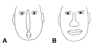
Fig. 1 Drawing of a face by the patient. A: Initially, she described the nose as looking very narrow as well as lengthened toward the mouth, which looked small and round of shape. B: She depicted a face normally to a certain degree at the time of discharge.

Fig. 2 A: Diffusion weighted MRI, and B: T2weighted brain MRI of the present patient, demonstrating a lesion in the fusiform face area. C: Fusiform face area. Viewed from bottom, the fusiform face area is depicted with blue.
Cited by 1 articles
-
Splenial Corpus Callosum Infarction Presenting with Unilateral Prosopometamorphopsia: A Case Report
Chang-Min Lee
Dement Neurocogn Disord. 2015;14(2):94-97. doi: 10.12779/dnd.2015.14.2.94.
Reference
-
1. Barton JJ, Press DZ, Keenan JP, O'Connor M. Lesions of the fusiform face area impair perception of facial configuration in prosopagnosia. Neurology. 2002. 58:71–78.
Article2. Dekowska M, Kuniecki M, Jaśkowski P. Facing facts: neuronal mechanisms of face perception. Acta Neurobiol Exp (Wars). 2008. 68:229–252.3. Bruce V, Young A. Understanding face recognition. Br J Psychol. 1986. 77:305–327.
Article4. Haxby JV, Hoffman EA, Gobbini MI. The distributed human neural system for face perception. Trends Cogn Sci. 2000. 4:223–233.
Article5. Kanwisher N, McDermott J, Chun MM. The fusiform face area: a module in human extrastriate cortex specialized for face perception. J Neurosci. 1997. 17:4302–4311.
Article6. Marotta JJ, Genovese CR, Behrmann M. A functional MRI study of face recognition in patients with prosopagnosia. Neuroreport. 2001. 12:1581–1587.
Article7. Miwa H, Kondo T. Metamorphopsia restricted to the right side of the face associated with a right temporal lobe lesion. J Neurol. 2007. 254:1765–1767.
Article
- Full Text Links
-
- Actions
-
Cited
- CITED
-
- Close
- Share
-
- Similar articles
-
- A Case of Prosopometamorphopsia Restricted to the Nose and Mouth with Right Medial Temporooccipital Lobe Infarction that Included the Fusiform Face Area
- Splenial Corpus Callosum Infarction Presenting with Unilateral Prosopometamorphopsia: A Case Report
- Prosopometamorphopsia and Visual Field Defect Improved by Valproic acid in a CADASIL Patient
- Strategic Infarct Dementia: Clinical Features, Neuroimaging and Neuropsychological Findings
- Consideration of Rescue Breathing methods during Infant Basic Life Support
