Dermoscopic Approach to a Small Round to Oval Hairless Patch on the Scalp
- Affiliations
-
- 1Department of Dermatology, Pusan National University School of Medicine, Busan, Korea. drkmp@hanmail.net
- 2Medical Research Institute, Pusan National University School of Medicine, Busan, Korea.
- KMID: 2171658
- DOI: http://doi.org/10.5021/ad.2014.26.2.214
Abstract
- BACKGROUND
Various kinds of alopecia can show small round or oval hairless patch. Dermoscopy could be a simple, useful tool for making a correct diagnosis.
OBJECTIVE
The aim of this study is to investigate clinical usefulness of dermoscopy for diseases with small round or oval hairless patch on the scalp.
METHODS
Dermoscopic examination was performed for 148 patients with small round or oval hairless patch using DermLite(R) II pro. The type and its patient number of alopecia investigated in the study were as below: alopecia areata (n=81), trichotillomania (n=24), tinea captis (n=13), traction alopecia (n=12), lichen planopilaris (n=8), discoid lupus erythematosus (n=7), congenital triangular alopecia (n=2) and pseudopelade of Brocq (n=1). The significance of dermoscopic findings for each disease were evaluated.
RESULTS
Characteristic dermoscopic findings of alopecia areata were tapering hairs and yellow dots. Those of trichotillomania and traction alopecia were broken hairs. Dermoscopic findings of tinea capitis included bent hairs, perifollicular white macules and greasy scales. Discoid lupus erythematosus and lichen planopilaris were characterized by dermoscopic findings of lack of follicular ostia. Furthermore, keratin plugs were frequently seen in discoid lupus erythematosus whereas perifollicular hyperkeratosis and erythema were frequently seen in lichen planopilaris.
CONCLUSION
Dermoscopic examination for small round or oval hairless patch showed characteristic findings for each disease. Based on these results, we propose dermoscopic algorithm for small round or oval hairless patch on the scalp.
Keyword
MeSH Terms
Figure
-
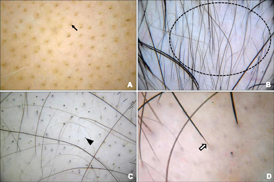
Fig. 1 Dermoscopic features of alopecia areata; yellow dots (A; arrow), clustered vellus hairs (B; dotted circle), black dots (C; triangle), and tapering hairs (D; white arrow).

Fig. 2 Dermoscopic features of trichotillomania; broken hairs and characteristic split end (arrow).
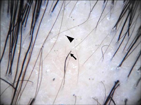
Fig. 3 Dermoscopic features of traction alopecia; broken hairs (arrow) and black dots (triangle).
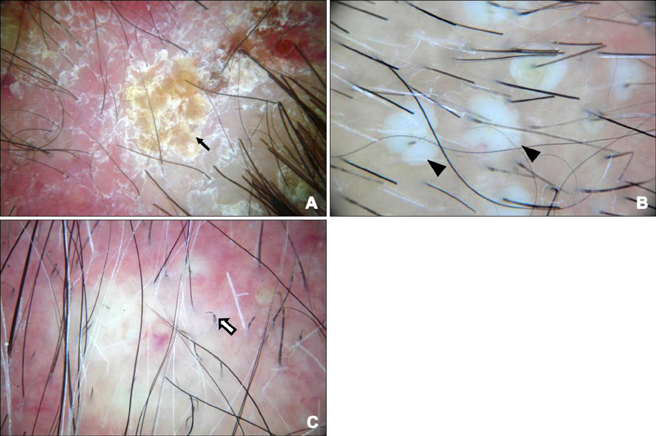
Fig. 4 Dermoscopic features of tinea capitis; greasy scales (A; arrow), perifollicular white macules (B; triangles) and bent hairs (C; white arrow).
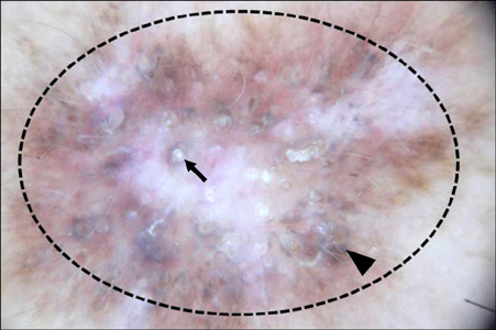
Fig. 5 Dermoscopic features of discoid lupus erythematosus; keratin plugs (arrow), lack of follicular ostia (dotted circle) and pigment network (triangle).
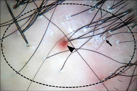
Fig. 6 Dermoscopic features of lichen planopilaris; perifollicular hyperkeratosis (arrow), perifollicularerythema (triangle) and lack of follicular ostia (dotted circle).
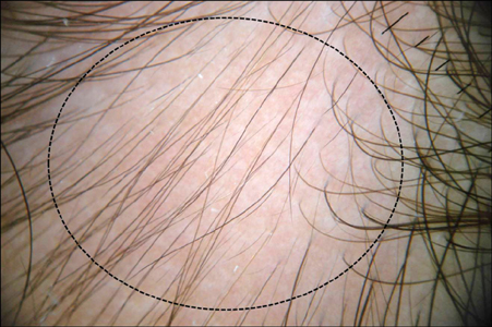
Fig. 7 Dermoscopic features of congenial triangular alopecia; clustered short vellus hairs without otherspecific findings (dotted circle).
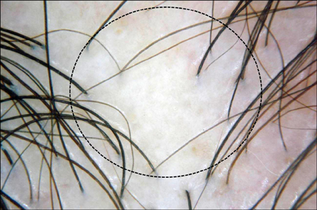
Fig. 8 Dermoscopic features of pseudopelade of Brocq; lack of follicular ostia (dotted circle) without otherspecific finding.

Fig. 9 The algorithm for dermoscopy of small round or oval hairless patch on the scalp. AA: alopecia areata, TM: trichotillomania, TA: traction alopecia, TC: tinea capitis, CTA: congenital triangular alopecia, DLE: discoid lupus erythematosus, LPP: lichen planopilaris, PB: pseudopelade of Brocq.
Cited by 1 articles
-
Trichoscopic Findings of Hair Loss in Koreans
Jin Park, Joo-Ik Kim, Han-Uk Kim, Seok-Kweon Yun, Seong-Jin Kim
Ann Dermatol. 2015;27(5):539-550. doi: 10.5021/ad.2015.27.5.539.
Reference
-
1. Inui S, Nakajima T, Nakagawa K, Itami S. Clinical significance of dermoscopy in alopecia areata: analysis of 300 cases. Int J Dermatol. 2008; 47:688–693.
Article2. Lee DY, Lee JH, Yang JM, Lee ES. The use of dermoscopy for the diagnosis of trichotillomania. J Eur Acad Dermatol Venereol. 2009; 23:731–732.
Article3. Lacarrubba F, Dall'Oglio F, Rita Nasca M, Micali G. Videodermatoscopy enhances diagnostic capability in some forms of hair loss. Am J Clin Dermatol. 2004; 5:205–208.
Article4. Slowinska M, Rudnicka L, Schwartz RA, Kowalska-Oledzka E, Rakowska A, Sicinska J, et al. Comma hairs: a dermatoscopic marker for tinea capitis: a rapid diagnostic method. J Am Acad Dermatol. 2008; 59:S77–S79.5. Duque-Estrada B, Tamler C, Sodré CT, Barcaui CB, Pereira FB. Dermoscopy patterns of cicatricial alopecia resulting from discoid lupus erythematosus and lichen planopilaris. An Bras Dermatol. 2010; 85:179–183.6. Wallace MP, de Berker DA. Hair diagnoses and signs: the use of dermatoscopy. Clin Exp Dermatol. 2010; 35:41–46.
Article7. Stefanato CM. Histopathology of alopecia: a clinicopathological approach to diagnosis. Histopathology. 2010; 56:24–38.
Article8. Fanti PA, Tosti A, Bardazzi F, Guerra L, Morelli R, Cameli N. Alopecia areata. A pathological study of nonresponder patients. Am J Dermatopathol. 1994; 16:167–170.9. Bielsa I, Herrero C, Collado A, Cobos A, Palou J, Mascaró JM. Histopathologic findings in cutaneous lupus erythematosus. Arch Dermatol. 1994; 130:54–58.
Article10. Kang H, Alzolibani AA, Otberg N, Shapiro J. Lichen planopilaris. Dermatol Ther. 2008; 21:249–256.
Article11. Isa-Isa R, Arenas R, Isa M. Inflammatory tinea capitis: kerion, dermatophytic granuloma, and mycetoma. Clin Dermatol. 2010; 28:133–136.
Article
- Full Text Links
-
- Actions
-
Cited
- CITED
-
- Close
- Share
-
- Similar articles
-
- Dermoscopic Approach to a Small Round to Oval Hairless Patch on the Scalp
- Correlation between Dermoscopic Features and Treatment Response in Discoid Lupus Erythematosus with Alopecia: A Retrospective Study
- Psychodynamic Approaches of Alopecia Totalis in Sisters
- A Case of Extramammary Paget's Disease on the Scalp
- Serially expanded flap use to treat large hairless scalp lesions
