J Korean Soc Spine Surg.
2001 Mar;8(1):39-45.
Posterior Lumbar Interbody Fusion in the Pyogenic Discitis
- Affiliations
-
- 1Department of Orthopaedic Surgery, College of Medicine, Pusan National University, Pusan, Korea. parkww@hyowon.pusan.ac.kr
Abstract
-
STUDY DESIGN: To present preliminary results of PLIF (Posterior lumbar interbody fusion) and pedicle screw fixation in the lum-bar pyogenic discitis.
OBJECTIVES
To evaluate the advantages and effects of PLIF and posterior instrumentation over recurrence of infection in lum-bar pyogenic discitis which are resistant to antibiotics. SUMMARY OF LITERATURE REVIEW: To the date, anterior removal of the focus followed by interposing autogenous bone graft without additional instrumentation and postoperative long-term immobilization has been the standard operative procedure.
MATERIALS AND METHODS
10 consecutive patients who had lumbar pyogenic discitis were treated by posterior approach from October 1997 to March 1999.
RESULTS
Based on MRI or CT finding, 9 solid union at 3-4 months after operation and 1 suspicious union at 1 year after opera-tion were observed. The mean preoperative lordotic angle of the affected segments was 9 degree compared to 20 degree after postoperation and 17 degree at last follow up. As for functional result of Kirkaldy-Willis, outcome was excellent in 3, good in 5, fair in 2, none poor case. The duration of postoperative bed rest period was an average of 3 days.
CONCLUSIONS
PLIF with instrumentation in lumbar pyogenic discitis is a useful treatment in posterior epidural abscess,coexis-tent spinal stenosis and lower lumbar level where anterior fixation is impossible. It is especially indicated in the case of scanty antevertebral abscess with minimal bone destruction. Its main advantage is early ambulation.
MeSH Terms
Figure
-
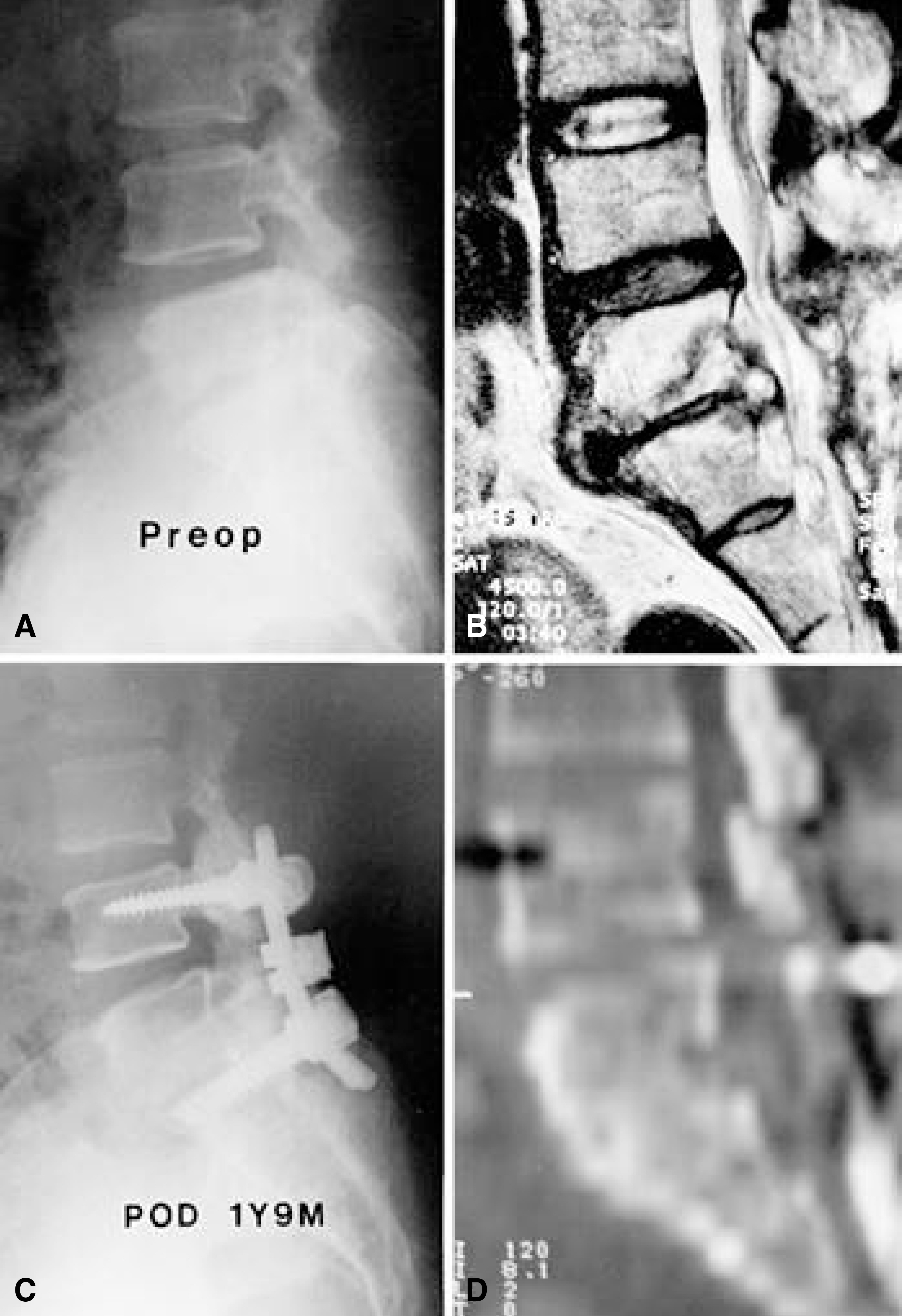
Fig. 1-A. Lateral radiographs shows L5-S1 disc space narrowing and irregular end plates. Fig. 1-B. T2-weighted image reveal a high intensity epidural abscess and destruction of the end plate. Als note the increased signal intensity in the involved surrounding vertebral bodies. Fig. 1-C. Follow-up lateral radiography, made 1 year 9 months after the operation, reveal solid fusion. Fig. 1-D. The CT scan demonstrates fusion between L5 and S1.
Reference
-
1). Costerton JW, Irvin RT and Cheng KJ. The bacterial glycocalyx in nature and disease. Ann Rev Microbiol. 35:299–324. 1981.
Article2). Eismont FJ, Bohlman HH, Soni PL, et al. Pyogenic and fungal vertebral osteomyelitis with paralysis. J Bone Joint Surg. 65-A:19–29. 1983.
Article3). Emery SE, Chan DP and Woodward HR. Treatment of hematogenous pyogenic vertebral osteomyelitis with Anterior debridement and primary bone grafting. Spine. 14:284–291. 1989.4). Eysel P, Hopf Ch, Vogel I and Rompe JD. Primary sta -ble anterior instrumentation or dorsoventral spondylode -sis in spondylodiscitis? Eur Spine J. 6:152–157. 1997.5). Fraser RD, Osti OL and Vernon-Roberts B. Iatrogenic discitis-the role of intravenous antibiotics in prevention and treatment: an experimental study. Spine. 14:1025–1032. 1989.6). Frederickson B, Yuan H and Olans R. Managemen t and outcome of pyogenic vertebral osteomyelitis. Clin Orthop. 131:160–167. 1978.7). Garcia A and Grantham SA. Hematogenous pyogenic vertebral osteomyelitis. J Bone Joint Surg. 42-A:429–436. 1960.8). J nsson B, S derholm R and Str mqvist B. Erythrocyte sedimentation rate after lumbar spine surgery. Spine. 16:1049–1050. 1991.
Article9). Kirkaldy-Willis WH, Paine KWE, Cauchoix J and Mclover G. Lumbar spinal stenosis. Clin Orthop. 99:30–52. 1974.10). Leung PC. Complication in the first 40 cases of microdiscectomy. J Spinal Dis. 1:306–310. 1988.11). Lindholm TS and Pylkkanen P. Discitis following removal of intervertebral disc. Spine. 7:618–622. 1982.
Article12). Medical Research Council Working Party on Tubercuosis of the Spine. A 10 year assessment of controlled trials of inpatient and outpatient treatment and of plaster-of- paris jackets for tuberculosis of the spine in children on standard chemotherapy. J Bone Joint Surg. 67-B:103–110. 1985.13). Oga M, Arizono T, Takasita M and Sugioka Y. Evaluation of the risk of instrumentation as a foreign body in spinal tuberculosis. Spine. 18:1890–1894. 1993.
Article14). Rajasekaran S and Shanmugasundaram TK. Prediction of the Angle of Gibbus Deformity in Tuberculosis of the Spine. J Bone Joint Surg. 69A:503–509. 1987.
- Full Text Links
-
- Actions
-
Cited
- CITED
-
- Close
- Share
-
- Similar articles
-
- Posterior Lumbar Interbody Fusion in the Pyogenic Discitis
- Posterior Lumbar Interbody Fusion Using Compressive Bone Graft with Allograft and Autograft in the Pyogenic Discitis
- Management of Lumbar Pyogenic Spondylitis with Posterior Decompression and Interbody Fusion Using Transpedicular Screws
- Anterior Lumbar Interbody Fusion with Percutaneous Pedicle Screw Fixation for the Treatment of Postoperative Pyogenic Spondylodiscitis
- One-Stage Posterior Debridement, Interbody Fusion and Instrumentation in the Treatment of Pyogenic Lumbar Spondylodiscitis
