J Korean Soc Transplant.
2010 Dec;24(4):311-315.
Clinical Efficacy of Pretransplant Magnetic Resonance Cholangiography of Donor for Living Donor Liver Transplantation
- Affiliations
-
- 1Department of Surgery, Sunchunhyang University Bucheon Hospital, Suncheonhyang University College of Medicine, Bucheon, Korea. hchulkim@schmc.ac.kr
Abstract
- BACKGROUND
Hepatobiliary and vascular structure anatomy must be understood to ensure donor safety during living donor liver transplantation (LDLT). The purpose of this study was to determine the role of pretransplant magnetic resonance cholangiography (MRC) for understanding the anatomy.
METHODS
Eighteen LDLT were analyzed retrospectively through medical records and radiological images. Pretransplant MRC and intraoperative cholangiography (IOC) were reviewed to evaluate the accuracy of pretransplant MRC.
RESULTS
The MRC results of 13 donors were acceptable for a living donor operation. However, 5 donor MRC results required further evaluation to identify the biliary anatomy by IOC. In 2 cases, the use of an intravenous low-dose morphine injection helped to obtain a more qualified MRC image.
CONCLUSIONS
Despite the small study size, the results showed that MRC can help provide information on donor biliary anatomy to ensure a safe donor operation.
Keyword
MeSH Terms
Figure
-
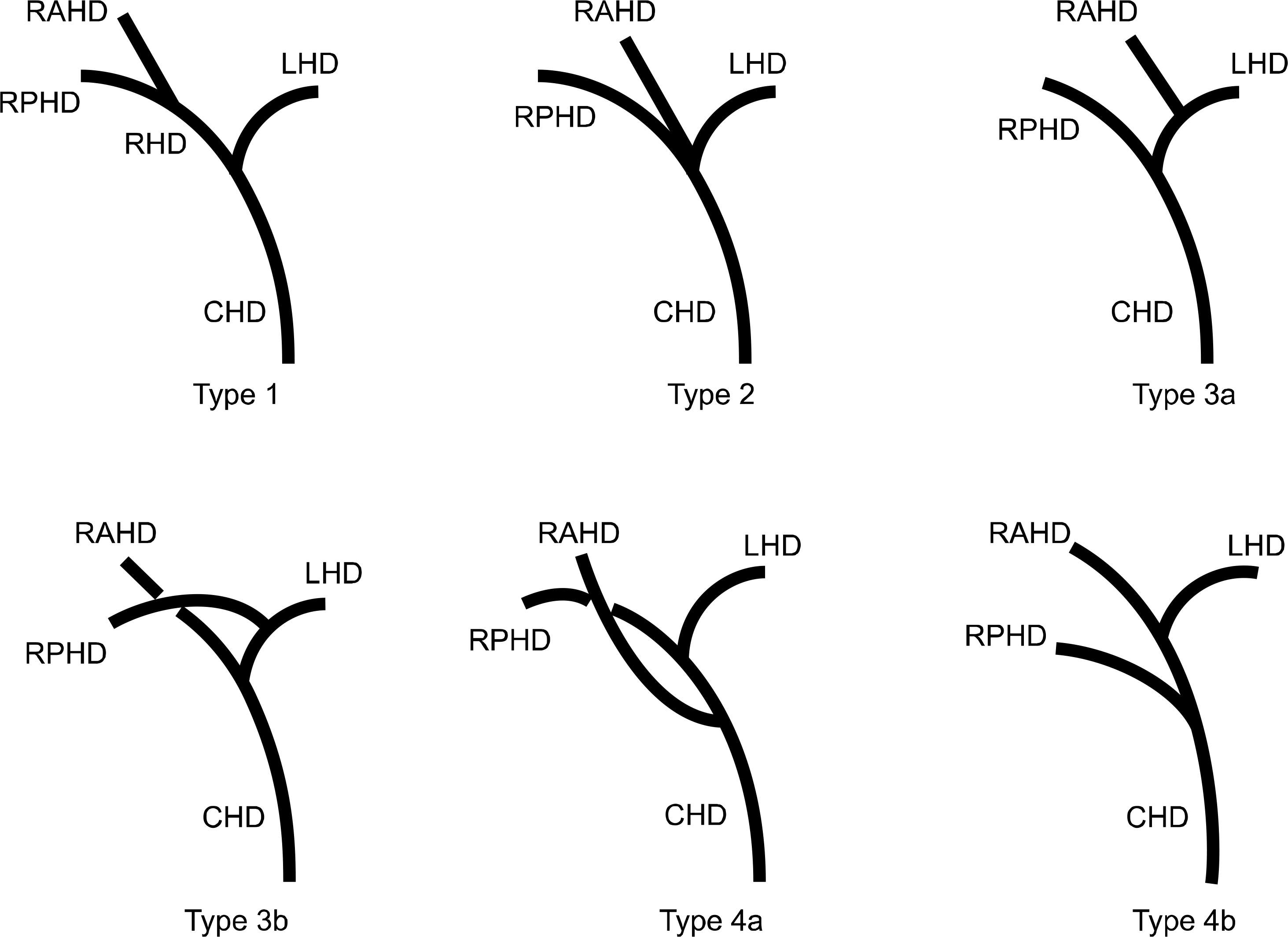
Fig. 1. Classification of the biliary tree anatomy. Abbreviations: CHD, common hepatic duct; LHD, left hepatic duct; RAHD, right anterior hepatic duct; RPHD, right posterior hepatic duct. Adapted from Fig. 3 of reference [20].
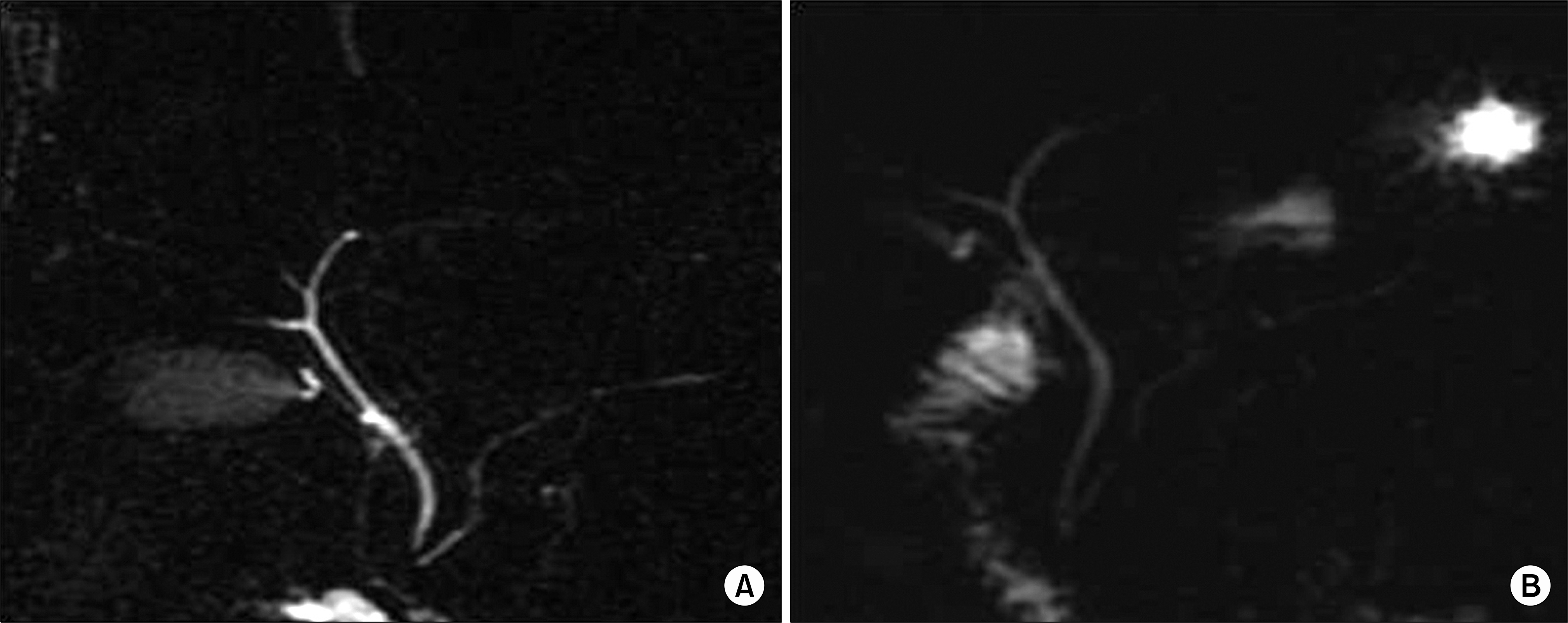
Fig. 2. Magnetic resonance cholangiopancreatographic image of a 21-year-old female following a morphine injection (A) and without a morphine injection in a 31-year-old man (B). The secondary bile duct and pancreatic duct are clearly visualized only in (A).
Reference
-
References
1). Korean Network for Organ Sharing (KONOS). Statistics of organ transplantation [Internet]. Seoul: KONOS;2009. Available from:. http://www.konos.go.kr.2). Pan GD, Yan LN. Problems in adult living donor liver transplantation using the right hepatic lobe. Hepatobiliary Pancreat Dis Int. 2006; 5:345–9.3). Hiroshige S, Shimada M, Harada N, Shiotani S, Ninomiya M, Minagawa R, et al. Accurate preoperative estimation of liver graft volumetry using three-dimensional computed tomography. Transplantation. 2003; 75:1561–4.4). Wallner BK, Schumacher KA, Weidenmaier W, Friedrich JM. Dilated biliary tract: evaluation with MR cholangiography with a T2-weighted contrast-enhanced fast sequence. Radiology. 1991; 181:805–8.
Article5). Hintze RE, Adler A, Veltzke W, Abou-Rebyeh H, Hammerstingl R, Vogl T. Clinical significance of magnetic resonance cholangiopancreatography (MRCP) compared to endosopic retrograde cholangiopancreatography (ERCP). Endoscopy. 1997; 29:182–7.6). Lim JS, Kim MJ, Myoung S, Park MS, Choi JY, Choi JS, et al. MR cholangiography for evaluation of hilar branching anatomy in transplantation of the right hepatic lobe from a living donor. AJR Am J Roentgenol. 2008; 191:537–45.
Article7). Ju MK, Kim MS, Choi GH, Chang HK, Ahn HJ, Kim YS, et al. The efficacy of pretransplant radiologic evaluation for graft volume and anatomy in living donor liver transplantation. J Korean Soc Transplant. 2007; 21:128–34. (주만기, 김명수, 최기홍, 장혜경, 안형준, 김유선, 등. 생체 간이식 공여자의 수술 전 이식간 용적과 해부학적 구조에 대한 영상학적 검사의 유용성. 대한이식학회지 2007;21: 128–34.).8). Irie H, Honda H, Kuroiwa T, Yoshimitsu K, Aibe H, Shinozaki K, et al. Pitfalls in MR cholangiopancreatographic interpretation. Radiographics. 2001; 21:23–37.
Article9). Thomas PD, Turner JG, Dobbs BR, Burt MJ, Chapman BA. Use of (99m)Tc-DISIDA biliary scanning with morphine provocation for the detection of elevated sphincter of Oddi basal pressure. Gut. 2000; 46:838–41.
Article10). Cappeliez O, Delhaye M, Devière J, Le Moine O, Metens T, Nicaise N, et al. Chronic pancreatitis: evaluation of pancreatic exocrine function with MR pancreatography after secretin stimulation. Radiology. 2000; 215:358–64.
Article11). Yamashita Y, Abe Y, Tang Y, Urata J, Sumi S, Takahashi M. In Vitro and clinical studies of image acquisition in breathhold MR cholangiopancreatography: single shot projection technique versus multislice technique. AJR Am J Roentgenol. 1997; 168:1449–54.12). Lee SJ, Ko JH, Cho YD, Jung MH, Yoon BC. Usefulness of MR cholangiopancreatography after intravenous morphine administration. J Korean Radiol Soc. 2007; 56:171–6. (고지호, 조영덕, 정미희, 윤병철. 자기공명담관이자조영술에서 정맥 내 모르핀 주입의 유용성. 대한영상의학회지 2007;56: 171–6.).
Article13). Silva AC, Friese JL, Hara AK, Liu PT. MR cholangiopancreatography: improved ductal distention with intravenous morphine administration. Radiographics. 2004; 24:677–87.
Article14). Nicaise N, Pellet O, Metens T, Devière J, Braudé P, Struyven J, et al. Magnetic resonance cholangiopancreatography: interest of IV secretin administration in the evaluation of pancreatic ducts. Eur Radiol. 1998; 8:16–22.
Article15). Helm JF, Venu RP, Geenen JE, Hogan WJ, Dodds WJ, Toouli J, et al. Effects of morphine on the human sphincter of Oddi. Gut. 1988; 29:1402–7.
Article16). Basaran C, Agildere AM, Donmez FY, Sevmis S, Budakoglu I, Karakayali H, et al. MR cholangiopancreatography with T2-weighted prospective acquisition correction turbo spinecho sequence of the biliary anatomy of potential living liver transplant donors. AJR Am J Roentgenol. 2008; 190:1527–33.
Article17). Goldman J, Florman S, Varotti G, Gondolesi GE, Gerning A, Fishbein T, et al. Noninvasive preoperative evaluation of biliary anatomy in right-lobe living donors with manga-fodipir trisodium-enhanced MR cholangiography. Transplant Proc. 2003; 35:1421–2.
Article18). Takatsuki M, Eguchi S, Yamanouchi K, Hidaka M, Soyama A, Kanematsu T. Technical refinements of bile duct division in living donor liver surgery. J Hepatobiliary Pancreat Sci [in press 2010 Aug 31].19). Taketomi A, Morita K, Toshima T, Takeishi K, Kayashima H, Ninomiya M, et al. Living donor hepatectomies with procedures to prevent biliary complications. J Am Coll Surg. 2010; 211:456–64.
Article20). Varotti G, Gondolesi GE, Goldman J, Wayne M, Florman SS, Schwartz ME, et al. Anatomic variations in right liver living donors. J Am Coll Surg. 2004; 198:577–82.
- Full Text Links
-
- Actions
-
Cited
- CITED
-
- Close
- Share
-
- Similar articles
-
- Clinical Efficacy of Pretransplant Magnetic Resonance Cholangiography of Donor for Living Donor Liver Transplantation
- The Role of Bile Duct Probe for Bile Duct Division during Donor Right Hemihepatectomy
- Pretransplant mycophenolate mofetil reduces intrahepatic cholangiopathy related to laparoscopic donor hepatectomy in ABO-incompatible liver transplantation
- Liver retransplantation for adult recipients
- Unilateral Versus Bilateral Biliary Drainage for Post-Transplant Anastomotic Stricture
