Cytological Results of Ultrasound-Guided Fine-Needle Aspiration Cytology for Thyroid Nodules: Emphasis on Correlation with Sonographic Findings
- Affiliations
-
- 1Department of Radiology and Research Institute of Radiological Science, Yonsei University College of Medicine, Seoul, Korea. ekkim@yuhs.ac
- 2Department of Pathology, Yonsei University College of Medicine, Seoul, Korea.
- 3Department of Surgery, Yonsei University College of Medicine, Seoul, Korea.
- KMID: 1108078
- DOI: http://doi.org/10.3349/ymj.2011.52.5.838
Abstract
- PURPOSE
To compare the cytological results of ultrasound-guided fine-needle aspiration (US-FNA) cytology of thyroid nodules to sonographic findings and determine whether US findings are helpful in the interpretation of cytological results.
MATERIALS AND METHODS
Among the thyroid nodules that underwent US-FNA cytology, we included the 819 nodules which had a conclusive diagnosis. Final diagnosis was based on pathology from surgery, repeated FNA cytology or follow-up of more than one year. Cytological results were divided into five groups: benign, indeterminate (follicular or Hurthle cell neoplasm), suspicious for malignancy, malignant, and inadequate. US findings were categorized as benign or suspicious. Cytological results and US categories were analyzed.
RESULTS
Final diagnosis was concluded upon in 819 nodules based on pathology (n=311), repeated FNA cytology (n=204) and follow-up (n=304), of which 634 were benign and 185 were malignant. There were 560 benign nodules, 141 malignant nodules, 49 nodules with inadequate results, 21 with indeterminate results, and 48 that were suspicious for malignancy. The positive and negative predictive values of the US categories were 59.1% and 97.0%, and those of the cytological results were 93.7% and 98.9%. The US categories were significantly correlated with final diagnosis in the benign (p=0.014) and suspicious for malignancy (p<0.001) cytological result groups, but not in the inadequate and indeterminate cytological results groups. The false positive and negative rates of cytological results were 1.9% and 3.2%.
CONCLUSION
Sonographic findings can be useful when used alongside cytological results, especially in nodules with cytological results that are benign or suspicious for malignancy.
MeSH Terms
Figure
-
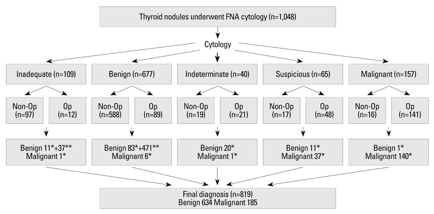
Fig. 1 Final diagnosis of the study population. FNA, fine-needle aspiration; Op, operation; NIC, no interval change. *from operation, **from repeated FNA cytology or NIC.
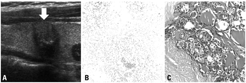
Fig. 2 A 50-year-old female with papillary thyroid carcinoma. (A) Ultrasonography shows well-circumscribed and isoechoic nodule (arrow) with a shape that is taller than it is wide and internal microcalcification in the thyroid gland; this nodule was considered suspicious. (B) Fine-needle aspiration cytology was interpreted as adenomatous hyperplasia based on the presence of flat sheets of follicular cells in the bloody background (Giemsa stain ×200 original magnification). Because of the discordant results between the sonographic findings and the cytological results, surgical excision was performed. (C) Thyroidectomy specimen shows a typical papillary carcinoma nucleus with a few grooves, clearing and pseudoinclusions, compatible with papillary thyroid carcinoma (hematoxylin and eosin (H&E) ×400 original magnification).
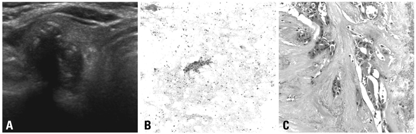
Fig. 3 A 61-year-old female with papillary thyroid carcinoma. (A) Ultrasonography shows an irregular and marked hypoechoic nodule with a shape that is taller than it is wide and internal mixed calcification in the right thyroid gland; this nodule was considered suspicious. (B) This fine-needle aspiration cytology was interpreted as suspicious for papillary thyroid carcinoma based on the presence of a rare cluster of follicular cells cytological overlapping and nuclear clearing and grooves (Papanicolaou stain ×200 original magnification). (C) This thyroidectomy specimen shows a few papillae with typical nuclear features of papillary carcinoma, such as nuclear pseudoinclusion, clearing and grooves, compatible with papillary carcinoma (H&E, ×400 original magnification).
Cited by 7 articles
-
Diagnostic Performance of Thyroglobulin Value in Indeterminate Range in Fine Needle Aspiration Washout Fluid from Lymph Nodes of Thyroid Cancer
Yu-Mee Sohn, Min Jung Kim, Eun-Kyung Kim, Jin Young Kwak
Yonsei Med J. 2012;53(1):126-131. doi: 10.3349/ymj.2012.53.1.126.Mixed Echoic Thyroid Nodules on Ultrasound: Approach to Management
Yu-Mee Sohn, Jung Hyun Yoon, Hee Jung Moon, Eun-Kyung Kim, Jin Young Kwak
Yonsei Med J. 2012;53(4):812-819. doi: 10.3349/ymj.2012.53.4.812.Anaplastic Thyroid Cancer: Ultrasonographic Findings and the Role of Ultrasonography-Guided Fine Needle Aspiration Biopsy
Hee Jung Suh, Hee Jung Moon, Jin Young Kwak, Ji Soo Choi, Eun-Kyung Kim
Yonsei Med J. 2013;54(6):1400-1406. doi: 10.3349/ymj.2013.54.6.1400.Diagnostic Role of Conventional Ultrasonography and Shearwave Elastography in Asymptomatic Patients with Diffuse Thyroid Disease: Initial Experience with 57 Patients
Injoong Kim, Eun-Kyung Kim, Jung Hyun Yoon, Kyung Hwa Han, Eun Ju Son, Hee Jung Moon, Jin Young Kwak
Yonsei Med J. 2014;55(1):247-253. doi: 10.3349/ymj.2014.55.1.247.High Body Mass Index and Thyroid Stimulating Hormone Levels Do Not Affect Thyroid Nodule Selection for Fine-Needle Aspiration Biopsy after Ultrasound Evaluation
Hyun Gi Kim, Hye Sun Lee, Eun Kyung Kim, Chung-Mo Nam, Hee Jung Moon, Hae Kyoung Jung, Jin Young Kwak
Int J Thyroidol. 2019;12(1):44-53. doi: 10.11106/ijt.2019.12.1.44.Value of Additional von Kossa Staining in Thyroid Nodules with “Suspicious for Malignancy” on Cytology
Hyeong Ju Kwon, Eun-Kyung Kim, Jin Young Kwak
J Korean Thyroid Assoc. 2015;8(1):81-87. doi: 10.11106/cet.2015.8.1.81.Usage and Diagnostic Yield of Fine-Needle Aspiration Cytology and Core Needle Biopsy in Thyroid Nodules: A Systematic Review and Meta-Analysis of Literature Published by Korean Authors
Soon-Hyun Ahn
Clin Exp Otorhinolaryngol. 2021;14(1):116-130. doi: 10.21053/ceo.2020.00199.
Reference
-
1. Nguyen GK, Lee MW, Ginsberg J, Wragg T, Bilodeau D. Fine-needle aspiration of the thyroid: an overview. Cytojournal. 2005. 2:12.2. Mazzaferri EL, de los Santos ET, Rofagha-Keyhani S. Solitary thyroid nodule: diagnosis and management. Med Clin North Am. 1988. 72:1177–1211.
Article3. Gharib H, Goellner JR. Fine-needle aspiration biopsy of the thyroid: an appraisal. Ann Intern Med. 1993. 118:282–289.
Article4. Kelly NP, Lim JC, DeJong S, Harmath C, Dudiak C, Wojcik EM. Specimen adequacy and diagnostic specificity of ultrasound-guided fine needle aspirations of nonpalpable thyroid nodules. Diagn Cytopathol. 2006. 34:188–190.
Article5. Cesur M, Corapcioglu D, Bulut S, Gursoy A, Yilmaz AE, Erdogan N, et al. Comparison of palpation-guided fine-needle aspiration biopsy to ultrasound-guided fine-needle aspiration biopsy in the evaluation of thyroid nodules. Thyroid. 2006. 16:555–561.
Article6. Khalid AN, Hollenbeak CS, Quraishi SA, Fan CY, Stack BC Jr. The cost-effectiveness of iodine 131 scintigraphy, ultrasonography, and fine-needle aspiration biopsy in the initial diagnosis of solitary thyroid nodules. Arch Otolaryngol Head Neck Surg. 2006. 132:244–250.
Article7. Bellantone R, Lombardi CP, Raffaelli M, Traini E, De Crea C, Rossi ED, et al. Management of cystic or predominantly cystic thyroid nodules: the role of ultrasound-guided fine-needle aspiration biopsy. Thyroid. 2004. 14:43–47.
Article8. Cai XJ, Valiyaparambath N, Nixon P, Waghorn A, Giles T, Helliwell T. Ultrasound-guided fine needle aspiration cytology in the diagnosis and management of thyroid nodules. Cytopathology. 2006. 17:251–256.
Article9. Yokozawa T, Fukata S, Kuma K, Matsuzuka F, Kobayashi A, Hirai K, et al. Thyroid cancer detected by ultrasound-guided fine-needle aspiration biopsy. World J Surg. 1996. 20:848–853.
Article10. Mittendorf EA, Tamarkin SW, McHenry CR. The results of ultrasound-guided fine-needle aspiration biopsy for evaluation of nodular thyroid disease. Surgery. 2002. 132:648–653.
Article11. Sangalli G, Serio G, Zampatti C, Bellotti M, Lomuscio G. Fine needle aspiration cytology of the thyroid: a comparison of 5469 cytological and final histological diagnoses. Cytopathology. 2006. 17:245–250.
Article12. Wu HH, Jones JN, Osman J. Fine-needle aspiration cytology of the thyroid: ten years experience in a community teaching hospital. Diagn Cytopathol. 2006. 34:93–96.
Article13. Kim EK, Park CS, Chung WY, Oh KK, Kim DI, Lee JT, et al. New sonographic criteria for recommending fine-needle aspiration biopsy of nonpalpable solid nodules of the thyroid. AJR Am J Roentgenol. 2002. 178:687–691.
Article14. Gharib H, Papini E, Valcavi R, Baskin HJ, Crescenzi A, Dottorini ME, et al. American Association of Clinical Endocrinologists and Associazione Medici Endocrinologi medical guidelines for clinical practice for the diagnosis and management of thyroid nodules. Endocr Pract. 2006. 12:63–102.15. Lee KY, Huang SM, Li S, Kim JM. Identification of differentially expressed genes in papillary thyroid cancers. Yonsei Med J. 2009. 50:60–67.
Article16. Akdi A, Perez G, Pastor S, Castell J, Biarnes J, Marcos R, et al. Common variants of the thyroglobulin gene are associated with differentiated thyroid cancer risk. Thyroid. 2011. 21:519–525.
Article17. Murugan AK, Xing M. Anaplastic Thyroid Cancers Harbor Novel Oncogenic Mutations of the ALK Gene. Cancer Res. 2011. 71:4403–4411.
Article18. Serna de la Saravia C, Cuellar F, Saravio Day E, Harach HR. Accuracy of aspiration cytology in thyroid cancer: a study in 1 institution. Acta Cytol. 2006. 50:384–387.19. Sahin M, Sengul A, Berki Z, Tutuncu NB, Guvener ND. Ultrasound-guided fine-needle aspiration biopsy and ultrasonographic features of infracentimetric nodules in patients with nodular goiter: correlation with pathological findings. Endocr Pathol. 2006. 17:67–74.
Article20. Zagorianakou P, Malamou-Mitsi V, Zagorianakou N, Stefanou D, Tsatsoulis A, Agnantis NJ. The role of fine-needle aspiration biopsy in the management of patients with thyroid nodules. In Vivo. 2005. 19:605–609.21. Sclabas GM, Staerkel GA, Shapiro SE, Fornage BD, Sherman SI, Vassillopoulou-Sellin R, et al. Fine-needle aspiration of the thyroid and correlation with histopathology in a contemporary series of 240 patients. Am J Surg. 2003. 186:702–709.
Article22. Ogawa Y, Kato Y, Ikeda K, Aya M, Ogisawa K, Kitani K, et al. The value of ultrasound-guided fine-needle aspiration cytology for thyroid nodules: an assessment of its diagnostic potential and pitfalls. Surg Today. 2001. 31:97–101.
Article23. Kessler A, Gavriel H, Zahav S, Vaiman M, Shlamkovitch N, Segal S, et al. Accuracy and consistency of fine-needle aspiration biopsy in the diagnosis and management of solitary thyroid nodules. Isr Med Assoc J. 2005. 7:371–373.24. Blanco Carrera C, Garcia-Diaz JD, Maqueda Villaizan E, Martinez-Onsurbe P, Pelaez Torres N, Saavedra Vallejo P. [Diagnostic efficacy of fine needle aspiration biopsy in patients with thyroid nodular disease. Analysis of 510 cases]. Rev Clin Esp. 2005. 205:374–378.25. Kim MJ, Kim EK, Park SI, Kim BM, Kwak JY, Kim SJ, et al. US-guided fine-needle aspiration of thyroid nodules: indications, techniques, results. Radiographics. 2008. 28:1869–1886.
Article26. Kwak JY, Kim EK, Kim MJ, Hong SW, Choi SH, Son EJ, et al. The role of ultrasound in thyroid nodules with a cytology reading of "suspicious for papillary thyroid carcinoma". Thyroid. 2008. 18:517–522.
Article27. Kwak JY, Kim EK, Kim HJ, Kim MJ, Son EJ, Moon HJ. How to combine ultrasound and cytological information in decision making about thyroid nodules. Eur Radiol. 2009. 19:1923–1931.
Article28. Moon HJ, Kwak JY, Kim EK, Kim MJ, Park CS, Chung WY, et al. The combined role of ultrasound and frozen section in surgical management of thyroid nodules read as suspicious for papillary thyroid carcinoma on fine needle aspiration biopsy: a retrospective study. World J Surg. 2009. 33:950–957.
Article29. BTA/RCP. Guidelines for the management of thyroid cancer. 2007. 2nd ed. London: The Lavenham Press.30. Baloch ZW, Fleisher S, LiVolsi VA, Gupta PK. Diagnosis of "follicular neoplasm": a gray zone in thyroid fine-needle aspiration cytology. Diagn Cytopathol. 2002. 26:41–44.
Article31. Rago T, Di Coscio G, Basolo F, Scutari M, Elisei R, Berti P, et al. Combined clinical, thyroid ultrasound and cytological features help to predict thyroid malignancy in follicular and Hupsilonrthle cell thyroid lesions: results from a series of 505 consecutive patients. Clin Endocrinol (Oxf). 2007. 66:13–20.32. Choi YJ, Yun JS, Kim DH. Clinical and ultrasound features of cytology diagnosed follicular neoplasm. Endocr J. 2009. 56:383–389.
Article33. Ylagan LR, Farkas T, Dehner LP. Fine needle aspiration of the thyroid: a cytohistologic correlation and study of discrepant cases. Thyroid. 2004. 14:35–41.
Article34. Carmeci C, Jeffrey RB, McDougall IR, Nowels KW, Weigel RJ. Ultrasound-guided fine-needle aspiration biopsy of thyroid masses. Thyroid. 1998. 8:283–289.
Article
- Full Text Links
-
- Actions
-
Cited
- CITED
-
- Close
- Share
-
- Similar articles
-
- Cytological Results of Ultrasound-Guided Fine-Needle Aspiration Cytology for Thyroid Nodules: Emphasis on Correlation with Sonographic Findings
- Diagnosis of Parathyroid Adenoma Detected during Thyroid Ultrasound: The Role of Parathormone Measurement in Fine-Needle Aspiration Washout
- Thyroid nodules with discordant results of ultrasonographic and fine-needle aspiration findings
- Follow-up of benign thyroid nodules confirmed by ultrasound-guided core needle biopsy after inconclusive cytology on fine-needle aspiration biopsy
- Ultrasound-Guided Fine-Needle Aspiration Biopsy of Multiple Thyroid Nodules
