Yonsei Med J.
2011 Sep;52(5):727-733. 10.3349/ymj.2011.52.5.727.
Application of Magnetic Resonance Imaging and Magnetic Resonance Angiography as Diagnostic Measures for the First Attack of Suspected Cerebrovascular Diseases in Korea
- Affiliations
-
- 1Department of Preventive Medicine, School of Medicine, Konkuk University, Seoul, Korea. mubul@kku.ac.kr
- 2Department of Neurology, Seoul Medical Center, Seoul, Korea.
- 3Department of Neurology, Seoul National University Bundang Hospital, College of Medicine, Seoul National University, Seongnam, Korea.
- 4Department of Neurology, National Medical Center, Seoul, Korea.
- KMID: 1108063
- DOI: http://doi.org/10.3349/ymj.2011.52.5.727
Abstract
- PURPOSE
No precise data are available showing how magnetic resonance imaging (MRI) and magnetic resonance angiography (MRA) can be applied to diagnosis for the first attack of a suspected cerebrovascular disease in Korea. The purpose of this study was to evaluate the application level of MRI and MRA as diagnostic tools and the related factors to the use of these techniques.
MATERIALS AND METHODS
This study used the health benefit claim data of 89,890 patients who were hospitalized for the first time due to suspected cerebrovascular disease in 2007 without having visited medical institutions as an outpatient or inpatient from 2003 to 2006.
RESULTS
Of the 89,890 cases, 28.4% took both MRI and MRA, 10.7% took only MRI and 6.9% took only MRA. The related factors identified in the multivariate logistic regression analysis were gender, type of insurance, type of medical institution, type of department, duration of hospitalization, and type of disease.
CONCLUSION
This study showed that the application level of MRI and MRA as diagnostic measures for the first attack of a suspected cerebrovascular diseases varied depending on several factors. It is necessary to study more accurate levels of computerized tomography (CT), computerized tomography angiography (CTA), MRI or MRA as measures to diagnose a first attack of suspected cerebrovascular disease.
Keyword
MeSH Terms
Figure
-
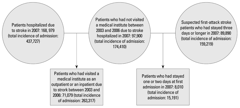
Fig. 1 Process of study subject selection.
Cited by 2 articles
-
Hemodynamic Significance of Internal Carotid or Middle Cerebral Artery Stenosis Detected on Magnetic Resonance Angiography
Hyo Jung Seo, Jefferson R. Pagsisihan, Jin Chul Paeng, Seung Hong Choi, Gi Jeong Cheon, June-Key Chung, Dong Soo Lee, Keon Wook Kang
Yonsei Med J. 2015;56(6):1686-1693. doi: 10.3349/ymj.2015.56.6.1686.The Brain MRI and MRA Findings of Patients Who Visited Memory Disorder Clinic in a General Hospital
Jun-Hyung Lee, Soo-Ji Lee, Jin-Young Ahn, Jae-Hyeok Heo
Dement Neurocogn Disord. 2012;11(4):124-130. doi: 10.12779/dnd.2012.11.4.124.
Reference
-
1. Korea National Statistical Office. Population Projections for Korea. 2006. Seoul: 1–56.2. Bae HJ. Epidemiology of stroke: 2006 Update. Korean J Stroke. 2007. 9:5–10.3. Rha JH. Stroke epidemiology 2007 Update. Korean J Stroke. 2008. 10:1–4.4. Bryan RN, Levy LM, Whitlow WD, Killian JM, Preziosi TJ, Rosario JA. Diagnosis of acute cerebral infarction: comparison of CT and MR imaging. AJNR Am J Neuroradiol. 1991. 12:611–620.5. Culebras A, Kase CS, Masdeu JC, Fox AJ, Bryan RN, Grossman CB, et al. Practice guidelines for the use of imaging in transient ischemic attacks and acute stroke. A report of the Stroke Council, American Heart Association. Stroke. 1997. 28:1480–1497.
Article6. Fiebach JB, Schellinger PD, Gass A, Kucinski T, Siebler M, Villringer A, et al. Stroke magnetic resonance imaging is accurate in hyperacute intracerebral hemorrhage: a multicenter study on the validity of stroke imaging. Stroke. 2004. 35:502–506.
Article7. Kidwell CS, Chalela JA, Saver JL, Starkman S, Hill MD, Demchuk AM, et al. Comparison of MRI and CT for detection of acute intracerebral hemorrhage. JAMA. 2004. 292:1823–1830.
Article8. Tsushima Y, Aoki J, Endo K. Brain microhemorrhages detected on T2*-weighted gradient-echo MR images. AJNR Am J Neuroradiol. 2003. 24:88–96.9. Dul K, Drayer BP. Kase CS, Caplan LR, editors. CT and MRI imaging of intracerebral hemorrhage. Intracerebral Hemorrhage. 1994. Boston, MA: Butterworth-Heinemann;73–93.10. Alvarez-Linera J, Benito-León J, Escribano J, Campollo J, Gesto R. Prospective evaluation of carotid artery stenosis: elliptic centric contrast-enhanced MR angiography and spiral CT angiography compared with digital subtraction angiography. AJNR Am J Neuroradiol. 2003. 24:1012–1019.11. Hirai T, Korogi Y, Ono K, Nagano M, Maruoka K, Uemura S, et al. Prospective evaluation of suspected stenoocclusive disease of the intracranial artery : combined MR angiogrphy and CT angiography compared with digital subtraction angiography. AJNR Am J Neuroradiol. 2002. 23:93–101.12. Skutta B, Fürst G, Eilers J, Ferbert A, Kuhn FP. Intracranial stenoocclusive disease: double-detector helical CT angiography versus digital subtraction angiography. AJNR Am J Neuroradiol. 1999. 20:791–799.13. Fazekas F, Niederkorn K, Ebner F, Díez-Tejedor E. Relevance of neuroimaging in the evaluation of cerebral ischemia. Cerebrovasc Dis. 2009. 27:Suppl 1. 1–8.
Article14. Korean Center for Disease Control and Prevention. Development of strategy for the role of community based cardio-cerebral disease center. 2009. Seoul:15. Korean Neurological Association. Role and future of neurology in geriatric medicine. 2009. Seoul:16. Wang L, Nie JX, Tracy CS, Moineddin R, Upshur RE. Utilization patterns of diagnostic imaging across the late life course: a population-based study in Ontario, Canada. Int J Technol Assess Health Care. 2008. 24:384–390.
Article17. Fowler RA, Sabur N, Li P, Juurlink DN, Pinto R, Hladunewich MA, et al. Sex-and age-based differences in the delivery and outcomes of critical care. CMAJ. 2007. 177:1513–1519.
Article18. Borkhoff CM, Hawker GA, Kreder HJ, Glazier RH, Mahomed NN, Wright JG. The effect of patients' sex on physicians' recommendations for total knee arthroplasty. CMAJ. 2008. 178:681–687.
Article19. Alter DA, Naylor CD, Austin PC, Chan BT, Tu JV. Geography and service supply do not explain socioeconomic gradients in angiography use after acute myocardial infarction. CMAJ. 2003. 168:261–264.20. Korean Center for Disease Control and Prevention. Development of strategy for the role of community based cardio-cerebral disease center. 2009. Seoul:21. Yoon SJ, Bae SC, Lee SI, Chang H, Jo H, Sung J, et al. Measuring the burden of disease in Korea. J Korean Med Sci. 2007. 22:518–523.
Article22. Demeter S, Reed M, Lix L, MacWilliam L, Leslie WD. Socioeconomic status and the utilization of diagnostic imaging in an urban setting. CMAJ. 2005. 173:1173–1177.
Article23. Adams HP Jr, Adams RJ, Brott T, del Zoppo GJ, Furlan A, Goldstein LB, et al. Guidelines for the early management of patients with ischemic stroke: A scientific statement from the Stroke Council of the American Stroke Association. Stroke. 2003. 34:1056–1083.
Article
- Full Text Links
-
- Actions
-
Cited
- CITED
-
- Close
- Share
-
- Similar articles
-
- Application of Magnetic Resonance Imaging and Magnetic Resonance Angiography as Diagnostic Measures for the First Attack of Suspected Cerebrovascular Diseases in Korea
- Functional Magnetic Resonance Imaging of the Brain: Principle and Practical Application
- Principles of Magnetic Resonance Angiography
- Bilateral Agenesis of the Internal Carotid Artery: Case Report
- Clinical application of high field strength magnetic resonance imaging
