Retrospective Electrocardiography-Gated Real-Time Cardiac Cine MRI at 3T: Comparison with Conventional Segmented Cine MRI
- Affiliations
-
- 1Department of Magnetic Resonance Imaging, Fuwai Hospital, State Key Laboratory of Cardiovascular Disease, National Center for Cardiovascular Diseases, Peking Union Medical College and Chinese Academy of Medical Sciences, Beijing, China. cjrzhaoshihua2009@163.com
- 2Cardiovascular Magnetic Resonance Unit, Royal Brompton Hospital, Sydney Street, London, England.
- KMID: 2429925
- DOI: http://doi.org/10.3348/kjr.2018.0243
Abstract
OBJECTIVE
Segmented cardiac cine magnetic resonance imaging (MRI) is the gold standard for cardiac ventricular volumetric assessment. In patients with difficulty in breath-holding or arrhythmia, this technique may generate images with inadequate quality for diagnosis. Real-time cardiac cine MRI has been developed to address this limitation. We aimed to assess the performance of retrospective electrocardiography-gated real-time cine MRI at 3T for left ventricular (LV) volume and mass measurement.
MATERIALS AND METHODS
Fifty-one patients were consecutively enrolled. A series of short-axis cine images covering the entire left ventricle using both segmented and real-time balanced steady-state free precession cardiac cine MRI were obtained. End-diastolic volume (EDV), end-systolic volume (ESV), stroke volume (SV), ejection fraction (EF), and LV mass were measured. The agreement and correlation of the parameters were assessed. Additionally, image quality was evaluated using European CMR Registry (Euro-CMR) score and structure visibility rating.
RESULTS
In patients without difficulty in breath-holding or arrhythmia, no significant difference was found in Euro-CMR score between the two techniques (0.3 ± 0.7 vs. 0.3 ± 0.5, p > 0.05). Good agreements and correlations were found between the techniques for measuring EDV, ESV, EF, SV, and LV mass. In patients with difficulty in breath-holding or arrhythmia, segmented cine MRI had a significant higher Euro-CMR score (2.3 ± 1.2 vs. 0.4 ± 0.5, p < 0.001).
CONCLUSION
Real-time cine MRI at 3T allowed the assessment of LV volume with high accuracy and showed a significantly better image quality compared to that of segmented cine MRI in patients with difficulty in breath-holding and arrhythmia.
MeSH Terms
Figure
-
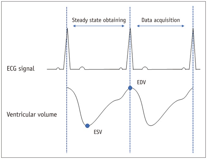
Fig. 1 Schematic diagram for data acquisition in retrospective gated real-time cardiac cine.ECG = electrocardiography, EDV = end-diastolic volume, ESV = end-systolic volume
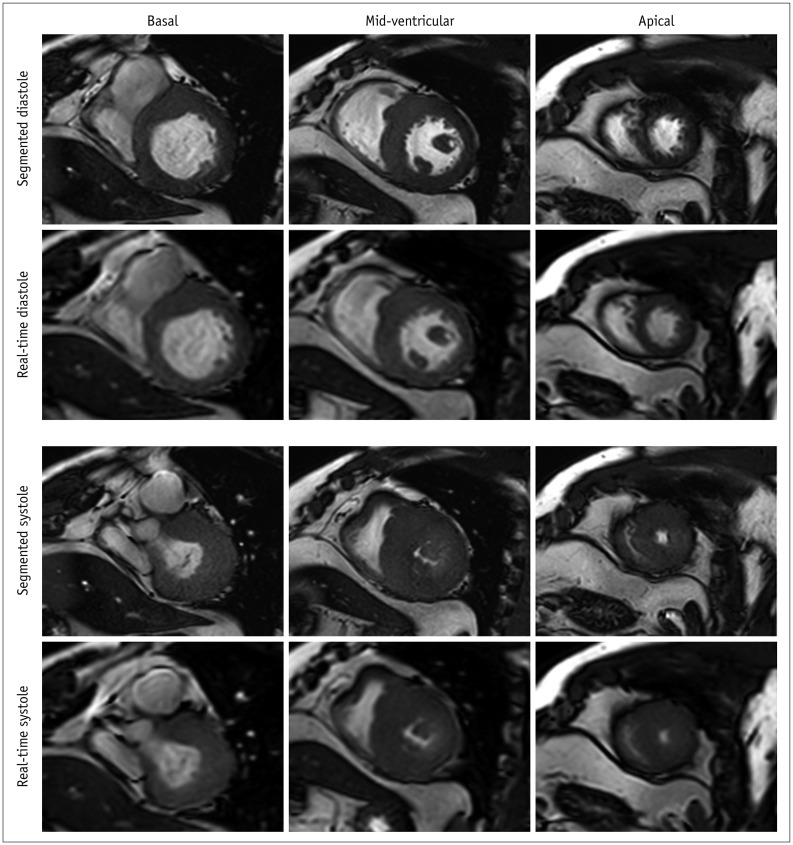
Fig. 2 Representative images of patients without difficulty in breath-holding and arrhythmia.Short-axis view images in end-diastole and end-systole phases in hypertrophic cardiomyopathy patient with heart rate of 59 beats/min.
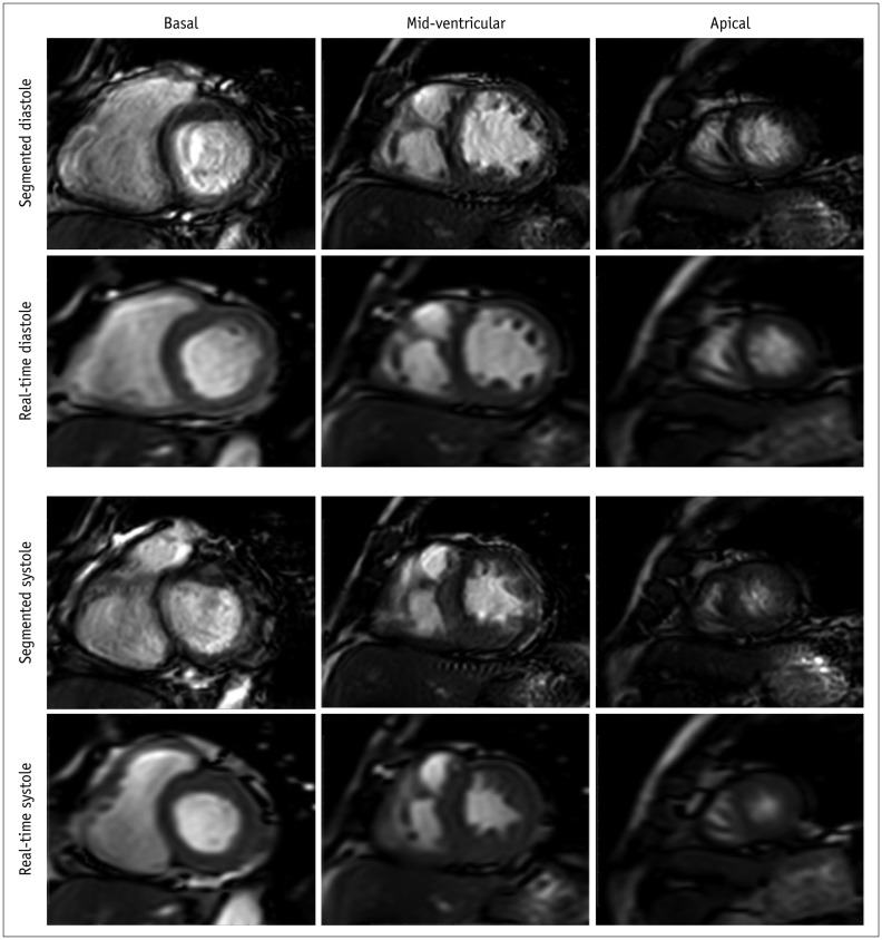
Fig. 3 Representative images of patients with difficulty in breath-holding and arrhythmia.Short-axis view images in end-diastole and end-systole phases in patient with atrial fibrillation. In contrast to real-time cardiac cine, endocardial contour was difficult to identify in conventional segmented cine in mid-ventricular and apical slices.

Fig. 4 Reviewer rating of endocardial border definition, epicardial border definition, papillary muscle visualization, myocardium visualization, blood pool contrast, and cardiac motion for real-time and segmented cine in patients from both groups.
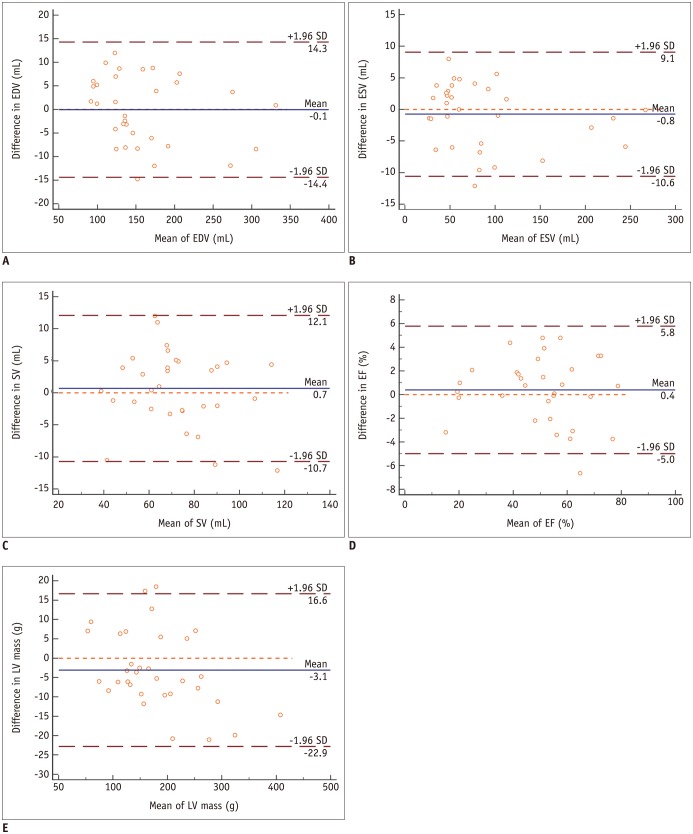
Fig. 5 Bland-Altman analysis shows good agreements in measurements of EDV (A), ESV (B), SV (C), EF (D) and LV mass (E).Blue line indicates mean value and brown dashed line indicates 95% confidence interval. EF = ejection fraction, LV = left ventricular, SD = standard deviation, SV = stroke volume
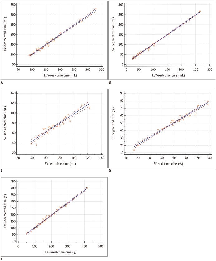
Fig. 6 Scatter plots for LV volumetric and mass measurements in standard segmented cine and real-time cine show good correlation in EDV (A), ESV (B), SV (C), EF (D), and mass (E) assessment.Brown dashed line indicates 95% confidence interval.
Cited by 2 articles
-
Guidelines for Cardiovascular Magnetic Resonance Imaging from the Korean Society of Cardiovascular Imaging—Part 2: Interpretation of Cine, Flow, and Angiography Data
Jae Wook Lee, Jee Hye Hur, Dong Hyun Yang, Bae Young Lee, Dong Jin Im, Su Jin Hong, Eun Young Kim, Eun-Ah Park, Yeseul Jo, JeongJae Kim, Chul Hwan Park, Hwan Seok Yong
Korean J Radiol. 2019;20(11):1477-1490. doi: 10.3348/kjr.2019.0407.Guideline for Cardiovascular Magnetic Resonance Imaging from the Korean Society of Cardiovascular Imaging—Part 1: Standardized Protocol
Yeseul Jo, JeongJae Kim, Chul Hwan Park, Jae Wook Lee, Jee Hye Hur, Dong Hyun Yang, Bae Young Lee, Dong Jin Im, Su Jin Hong, Eun Young Kim, Eun-Ah Park, Pan Ki Kim, Hwan Seok Yong
Korean J Radiol. 2019;20(9):1313-1333. doi: 10.3348/kjr.2019.0398.
Reference
-
1. Knauth AL, Gauvreau K, Powell AJ, Landzberg MJ, Walsh EP, Lock JE, et al. Ventricular size and function assessed by cardiac MRI predict major adverse clinical outcomes late after tetralogy of Fallot repair. Heart. 2008; 94:211–216. PMID: 17135219.
Article2. Curtis JP, Sokol SI, Wang Y, Rathore SS, Ko DT, Jadbabaie F, et al. The association of left ventricular ejection fraction, mortality, and cause of death in stable outpatients with heart failure. J Am Coll Cardiol. 2003; 42:736–742. PMID: 12932612.
Article3. Karamitsos TD, Francis JM, Myerson S, Selvanayagam JB, Neubauer S. The role of cardiovascular magnetic resonance imaging in heart failure. J Am Coll Cardiol. 2009; 54:1407–1424. PMID: 19796734.
Article4. Moon JC, Lorenz CH, Francis JM, Smith GC, Pennell DJ. Breath-hold FLASH and FISP cardiovascular MR imaging: left ventricular volume differences and reproducibility. Radiology. 2002; 223:789–797. PMID: 12034951.
Article5. Kühl HP, Spuentrup E, Wall A, Franke A, Schröder J, Heussen N, et al. Assessment of myocardial function with interactive non-breath-hold real-time MR imaging: comparison with echocardiography and breath-hold cine MR imaging. Radiology. 2004; 231:198–207. PMID: 14990805.
Article6. Hori Y, Yamada N, Higashi M, Hirai N, Nakatani S. Rapid evaluation of right and left ventricular function and mass using real-time true-FISP cine MR imaging without breath-hold: comparison with segmented true-FISP cine MR imaging with breath-hold. J Cardiovasc Magn Reson. 2003; 5:439–450. PMID: 12882075.
Article7. Voit D, Zhang S, Unterberg-Buchwald C, Sohns JM, Lotz J, Frahm J. Real-time cardiovascular magnetic resonance at 1.5 T using balanced SSFP and 40 ms resolution. J Cardiovasc Magn Reson. 2013; 15:79. PMID: 24028285.
Article8. Kaji S, Yang PC, Kerr AB, Tang WH, Meyer CH, Macovski A, et al. Rapid evaluation of left ventricular volume and mass without breath-holding using real-time interactive cardiac magnetic resonance imaging system. J Am Coll Cardiol. 2001; 38:527–533. PMID: 11499748.
Article9. Schwab F, Schwarz F, Dietrich O, Lanz T, Resmer F, Wichmann T, et al. Free breathing real-time cardiac cine imaging with improved spatial resolution at 3 T. Invest Radiol. 2013; 48:158–166. PMID: 23344515.
Article10. Sudarski S, Henzler T, Haubenreisser H, Dösch C, Zenge MO, Schmidt M, et al. Free-breathing sparse sampling cine MR imaging with iterative reconstruction for the assessment of left ventricular function and mass at 3.0 T. Radiology. 2017; 282:74–83. PMID: 27399326.11. Vincenti G, Monney P, Chaptinel J, Rutz T, Coppo S, Zenge MO, et al. Compressed sensing single-breath-hold CMR for fast quantification of LV function, volumes, and mass. JACC Cardiovasc Imaging. 2014; 7:882–892. PMID: 25129517.12. Yamamuro M, Tadamura E, Kanao S, Okayama S, Okamoto J, Urayama S, et al. Cardiac functional analysis by free-breath real-time cine CMR with a spatiotemporal filtering method, TSENSE: comparison with breath-hold cine CMR. J Cardiovasc Magn Reson. 2006; 8:801–807. PMID: 17060102.
Article13. Feng L, Srichai MB, Lim RP, Harrison A, King W, Adluru G, et al. Highly accelerated real-time cardiac cine MRI using k-t SPARSE-SENSE. Magn Reson Med. 2013; 70:64–74. PMID: 22887290.14. Xue H, Kellman P, Larocca G, Arai AE, Hansen MS. High spatial and temporal resolution retrospective cine cardiovascular magnetic resonance from shortened free breathing real-time acquisitions. J Cardiovasc Magn Reson. 2013; 15:102. PMID: 24228930.
Article15. Kellman P, Chefd'hotel C, Lorenz CH, Mancini C, Arai AE, McVeigh ER. Fully automatic, retrospective enhancement of real-time acquired cardiac cine MR images using image-based navigators and respiratory motion-corrected averaging. Magn Reson Med. 2008; 59:771–778. PMID: 18302227.
Article16. Sievers B, Kirchberg S, Bakan A, Franken U, Trappe HJ. Impact of papillary muscles in ventricular volume and ejection fraction assessment by cardiovascular magnetic resonance. J Cardiovasc Magn Reson. 2004; 6:9–16. PMID: 15054924.
Article17. Klinke V, Muzzarelli S, Lauriers N, Locca D, Vincenti G, Monney P, et al. Quality assessment of cardiovascular magnetic resonance in the setting of the European CMR registry: description and validation of standardized criteria. J Cardiovasc Magn Reson. 2013; 15:55. PMID: 23787094.
Article18. Camargo GC, Erthal F, Sabioni L, Penna F, Strecker R, Schmidt M, et al. Real-time cardiac magnetic resonance cine imaging with sparse sampling and iterative reconstruction for left-ventricular measures: comparison with gold-standard segmented steady-state free precession. Magn Reson Imaging. 2017; 38:138–144. PMID: 28065694.
Article19. Aandal G, Nadig V, Yeh V, Rajiah P, Jenkins T, Sattar A, et al. Evaluation of left ventricular ejection fraction using through-time radial GRAPPA. J Cardiovasc Magn Reson. 2014; 16:79. PMID: 25315256.
Article20. Siontis KC, Geske JB, Ong K, Nishimura RA, Ommen SR, Gersh BJ. Atrial fibrillation in hypertrophic cardiomyopathy: prevalence, clinical correlations, and mortality in a large high-risk population. J Am Heart Assoc. 2014; 3:e001002. PMID: 24965028.
Article21. Ceresne L, Upshur RE. Atrial fibrillation in a primary care practice: prevalence and management. BMC Fam Pract. 2002; 3:11. PMID: 12031095.
Article
- Full Text Links
-
- Actions
-
Cited
- CITED
-
- Close
- Share
-
- Similar articles
-
- Retrospective Electrocardiography-Gated Real-Time Cardiac Cine MRI at 3T: Comparison with Conventional Segmented Cine MRI
- Comparison between Echocardiography and Cardiac Cine-MRI : Left Ventricular Volume and Cardiac Output
- Comparison of Left and Right Ventricular Volume and Cardiac Output by MRI and Echocardiography
- Comparison between Three-Dimensional Navigator-Gated Whole-Heart MRI and Two-Dimensional Cine MRI in Quantifying Ventricular Volumes
- Comparison of Cine Magnetic Resonance Imaging with Doppler Echocardiography for the Quantative Evaluation of Tricuspid Regurgitation in Newborn
