Selection and Reporting of Statistical Methods to Assess Reliability of a Diagnostic Test: Conformity to Recommended Methods in a Peer-Reviewed Journal
- Affiliations
-
- 1Department of Radiology and Research Institute of Radiology, University of Ulsan College of Medicine, Asan Medical Center, Seoul 05505, Korea. parksh.radiology@gmail.com
- 2Department of Radiology, Research Institute of Radiological Science, Yonsei University College of Medicine, Seoul 03722, Korea.
- 3Department of Radiology, Chung-Ang University Hospital, Chung-Ang University College of Medicine, Seoul 06973, Korea.
- 4Korea Research Institute of Standards and Science, Daejeon 34113, Korea.
- KMID: 2427198
- DOI: http://doi.org/10.3348/kjr.2017.18.6.888
Abstract
OBJECTIVE
To evaluate the frequency and adequacy of statistical analyses in a general radiology journal when reporting a reliability analysis for a diagnostic test.
MATERIALS AND METHODS
Sixty-three studies of diagnostic test accuracy (DTA) and 36 studies reporting reliability analyses published in the Korean Journal of Radiology between 2012 and 2016 were analyzed. Studies were judged using the methodological guidelines of the Radiological Society of North America-Quantitative Imaging Biomarkers Alliance (RSNA-QIBA), and COnsensus-based Standards for the selection of health Measurement INstruments (COSMIN) initiative. DTA studies were evaluated by nine editorial board members of the journal. Reliability studies were evaluated by study reviewers experienced with reliability analysis.
RESULTS
Thirty-one (49.2%) of the 63 DTA studies did not include a reliability analysis when deemed necessary. Among the 36 reliability studies, proper statistical methods were used in all (5/5) studies dealing with dichotomous/nominal data, 46.7% (7/15) of studies dealing with ordinal data, and 95.2% (20/21) of studies dealing with continuous data. Statistical methods were described in sufficient detail regarding weighted kappa in 28.6% (2/7) of studies and regarding the model and assumptions of intraclass correlation coefficient in 35.3% (6/17) and 29.4% (5/17) of studies, respectively. Reliability parameters were used as if they were agreement parameters in 23.1% (3/13) of studies. Reproducibility and repeatability were used incorrectly in 20% (3/15) of studies.
CONCLUSION
Greater attention to the importance of reporting reliability, thorough description of the related statistical methods, efforts not to neglect agreement parameters, and better use of relevant terminology is necessary.
Keyword
MeSH Terms
Figure
-
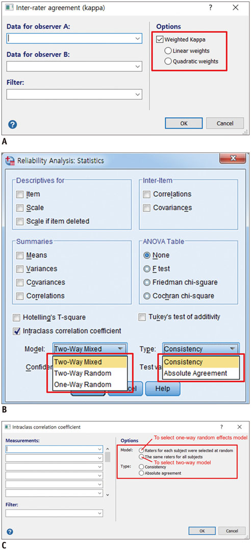
Fig. 1 Display of detailed options associated with statistical tests used for reliability analysis in some user-friendly software programs. A. Selection of weighting method to calculate weighted kappa with MedCalc Version 17.6 (MedCalc Software BVBA; https://www.medcalc.org). B. Selection of model and assumption to calculate ICC with IBM SPSS Statistics for Windows Version 21 (IBM Corp.). C. Selection of model and assumption to calculate ICC with MedCalc Version 17.6 (MedCalc Software BVBA). This software program does not distinguish between random and fixed effects models. ICC = intraclass correlation coefficient
Cited by 4 articles
-
Optimal Phase of Dynamic Computed Tomography for Reliable Size Measurement of Metastatic Neuroendocrine Tumors of the Liver: Comparison between Pre- and Post-Contrast Phases
Jimi Huh, Jisuk Park, Kyung Won Kim, Hyoung Jung Kim, Jong Seok Lee, Jong Hwa Lee, Yoong Ki Jeong, Atul B. Shinagare, Nikhil H. Ramaiya
Korean J Radiol. 2018;19(6):1066-1076. doi: 10.3348/kjr.2018.19.6.1066.Age of Data in Contemporary Research Articles Published in Representative General Radiology Journals
Ji Hun Kang, Dong Hwan Kim, Seong Ho Park, Jung Hwan Baek
Korean J Radiol. 2018;19(6):1172-1178. doi: 10.3348/kjr.2018.19.6.1172.Prognostic Value of Radiologic Extranodal Extension in Human Papillomavirus-Related Oropharyngeal Squamous Cell Carcinoma
Boeun Lee, Young Jun Choi, Seon-Ok Kim, Yoon Se Lee, Jung Yong Hong, Jung Hwan Baek, Jeong Hyun Lee
Korean J Radiol. 2019;20(8):1266-1274. doi: 10.3348/kjr.2018.0742.High-Resolution Magnetic Resonance Imaging Using Compressed Sensing for Intracranial and Extracranial Arteries: Comparison with Conventional Parallel Imaging
Chong Hyun Suh, Seung Chai Jung, Ho Beom Lee, Se Jin Cho
Korean J Radiol. 2019;20(3):487-497. doi: 10.3348/kjr.2018.0424.
Reference
-
1. Bankier AA, Levine D, Halpern EF, Kressel HY. Consensus interpretation in imaging research: is there a better way? Radiology. 2010; 257:14–17.2. Reid MC, Lachs MS, Feinstein AR. Use of methodological standards in diagnostic test research. Getting better but still not good. JAMA. 1995; 274:645–651.3. Levine D, Bankier AA, Halpern EF. Submissions to radiology: our top 10 list of statistical errors. Radiology. 2009; 253:288–290.4. Hernaez R. Reliability and agreement studies: a guide for clinical investigators. Gut. 2015; 64:1018–1027.5. Kessler LG, Barnhart HX, Buckler AJ, Choudhury KR, Kondratovich MV, Toledano A, et al. The emerging science of quantitative imaging biomarkers terminology and definitions for scientific studies and regulatory submissions. Stat Methods Med Res. 2015; 24:9–26.6. Raunig DL, McShane LM, Pennello G, Gatsonis C, Carson PL, Voyvodic JT, et al. Quantitative imaging biomarkers: a review of statistical methods for technical performance assessment. Stat Methods Med Res. 2015; 24:27–67.7. Kottner J, Audigé L, Brorson S, Donner A, Gajewski BJ, Hróbjartsson A, et al. Guidelines for reporting reliability and agreement Studies (GRRAS) were proposed. J Clin Epidemiol. 2011; 64:96–106.8. Bossuyt PM, Reitsma JB, Bruns DE, Gatsonis CA, Glasziou PP, Irwig L, et al. STARD 2015: an updated list of essential items for reporting diagnostic accuracy studies. Radiology. 2015; 277:826–832.9. Choi YJ, Chung MS, Koo HJ, Park JE, Yoon HM, Park SH. Does the reporting quality of diagnostic test accuracy studies, as defined by STARD 2015, affect citation? Korean J Radiol. 2016; 17:706–714.10. Gallo L, Hua N, Mercuri M, Silveira A, Worster A. Best Evidence in Emergency Medicine (BEEM; beem.ca). Adherence to standards for reporting diagnostic accuracy in emergency medicine research. Acad Emerg Med. 2017; 06. 16. [Epub]. DOI: 10.1111/acem.13233.11. Grob AT, van der Vaart LR, Withagen MI, van der Vaart CH. The quality of reporting of diagnostic accuracy studies in pelvic floor transperineal three-dimensional ultrasound: a systematic review. Ultrasound Obstet Gynecol. 2016; 12. 21. [Epub]. DOI: 10.1002/uog.17390.12. Hong PJ, Korevaar DA, McGrath TA, Ziai H, Frank R, Alabousi M, et al. Reporting of imaging diagnostic accuracy studies with focus on MRI subgroup: Adherence to STARD 2015. J Magn Reson Imaging. 2017; 06. 22. [Epub]. DOI: 10.1002/jmri.25797.13. Korevaar DA, Cohen JF, Hooft L, Bossuyt PM. Literature survey of high-impact journals revealed reporting weaknesses in abstracts of diagnostic accuracy studies. J Clin Epidemiol. 2015; 68:708–715.14. Korevaar DA, Wang J, van Enst WA, Leeflang MM, Hooft L, Smidt N, et al. Reporting diagnostic accuracy studies: some improvements after 10 years of STARD. Radiology. 2015; 274:781–789.15. Xu XQ, Hu H, Su GY, Liu H, Shi HB, Wu FY. Diffusion weighted imaging for differentiating benign from malignant orbital tumors: diagnostic performance of the apparent diffusion coefficient based on region of interest selection method. Korean J Radiol. 2016; 17:650–656.16. Park HJ, Lee SY, Rho MH, Chung EC, Kim MS, Kwon HJ, et al. Single-shot echo-planar diffusion-weighted MR imaging at 3T and 1.5T for differentiation of benign vertebral fracture edema and tumor infiltration. Korean J Radiol. 2016; 17:590–597.17. Liu J, Ji Y, Ai H, Ning B, Zhao J, Zhang Y, et al. Liver shear-wave velocity and serum fibrosis markers to diagnose hepatic fibrosis in patients with chronic viral hepatitis B. Korean J Radiol. 2016; 17:396–404.18. Lim HJ, Chung MJ, Shin KE, Hwang HS, Lee KS. The impact of iterative reconstruction in low-dose computed tomography on the evaluation of diffuse interstitial lung disease. Korean J Radiol. 2016; 17:950–960.19. Lee JE, Cho JS, Shin KS, Kim SS, You SK, Park JW, et al. Diffuse infiltrative splenic lymphoma: diagnostic efficacy of arterial-phase CT. Korean J Radiol. 2016; 17:734–741.20. Lee H, Lee WW, Park SY, Kim SE. F-18 sodium fluoride positron emission tomography/computed tomography for detection of thyroid cancer bone metastasis compared with bone scintigraphy. Korean J Radiol. 2016; 17:281–288.21. Lee EH, Kim KW, Kim YJ, Shin DR, Park YM, Lim HS, et al. Performance of screening mammography: a report of the alliance for breast cancer screening in Korea. Korean J Radiol. 2016; 17:489–496.22. Joo SM, Kim YP, Yum TJ, Eun NL, Lee D, Lee KH. Optimized performance of flightplan during chemoembolization for hepatocellular carcinoma: importance of the proportion of segmented tumor area. Korean J Radiol. 2016; 17:771–778.23. Hong HS, Cho HS, Woo JY, Lee Y, Yang I, Hwang JY, et al. Intra-appendiceal air at CT: is it a useful or a confusing sign for the diagnosis of acute appendicitis? Korean J Radiol. 2016; 17:39–46.24. Ha EJ, Moon WJ, Na DG, Lee YH, Choi N, Kim SJ, et al. A multicenter prospective validation study for the Korean thyroid imaging reporting and data system in patients with thyroid nodules. Korean J Radiol. 2016; 17:811–821.25. Ding Y, Zeng M, Rao S, Chen C, Fu C, Zhou J. Comparison of biexponential and monoexponential model of diffusion-weighted imaging for distinguishing between common renal cell carcinoma and fat poor angiomyolipoma. Korean J Radiol. 2016; 17:853–863.26. Choi TW, Lee JM, Kim JH, Yu MH, Han JK, Choi BI. Comparison of multidetector CT and gadobutrol-enhanced MR imaging for evaluation of small, solid pancreatic lesions. Korean J Radiol. 2016; 17:509–521.27. Alan B, Göya C, Tunç S, Teke M, Hattapoğlu S. Assessment of placental stiffness using acoustic radiation force impulse elastography in pregnant women with fetal anomalies. Korean J Radiol. 2016; 17:218–223.28. Ahn JH, Yu JS, Cho ES, Chung JJ, Kim JH, Kim KW. Diffusion-weighted MRI of malignant versus benign portal vein thrombosis. Korean J Radiol. 2016; 17:533–540.29. Ryoo I, Suh S, Lee YH, Seo HS, Seol HY. Comparison of ultrasonographic findings of biopsy-proven tuberculous lymphadenitis and kikuchi disease. Korean J Radiol. 2015; 16:767–775.30. Park B, Kim HK, Choi YS, Kim J, Zo JI, Choi JY, et al. Prediction of pathologic grade and prognosis in mucoepidermoid carcinoma of the lung using 18F-FDG PET/CT. Korean J Radiol. 2015; 16:929–935.31. Niu XK, Bhetuwal A, Yang HF. CT-guided core needle biopsy of pleural lesions: evaluating diagnostic yield and associated complications. Korean J Radiol. 2015; 16:206–212.32. Lu Q, Huang BJ, Wang WP, Li CX, Xue LY. Qualitative and quantitative analysis with contrast-enhanced ultrasonography: diagnosis value in hypoechoic renal angiomyolipoma. Korean J Radiol. 2015; 16:334–341.33. Lee S, Lee YH, Chung TS, Jeong EK, Kim S, Yoo YH, et al. Accuracy of diffusion tensor imaging for diagnosing cervical spondylotic myelopathy in patients showing spinal cord compression. Korean J Radiol. 2015; 16:1303–1312.34. Lee EK, Choi SH, Yun TJ, Kang KM, Kim TM, Lee SH, et al. Prediction of response to concurrent chemoradiotherapy with temozolomide in glioblastoma: application of immediate post-operative dynamic susceptibility contrast and diffusion-weighted MR imaging. Korean J Radiol. 2015; 16:1341–1348.35. Kim SA, Chang JM, Cho N, Yi A, Moon WK. Characterization of breast lesions: comparison of digital breast tomosynthesis and ultrasonography. Korean J Radiol. 2015; 16:229–238.36. Kim J, Kim YH, Lee KH, Lee YJ, Park JH. Diagnostic performance of CT angiography in patients visiting emergency department with overt gastrointestinal bleeding. Korean J Radiol. 2015; 16:541–549.37. Kim DW, Jung SL, Kim J, Ryu JH, Sung JY, Lim HK. Comparison between ultrasonography and computed tomography for detecting the pyramidal lobe of the thyroid gland: a prospective multicenter study. Korean J Radiol. 2015; 16:402–409.38. Kang KA, Kim YK, Kim E, Jeong WK, Choi D, Lee WJ, et al. T2-weighted liver MRI using the multivane technique at 3T: comparison with conventional T2-weighted MRI. Korean J Radiol. 2015; 16:1038–1046.39. Jung SI, Park HS, Yim Y, Jeon HJ, Yu MH, Kim YJ, et al. Added value of using a CT coronal reformation to diagnose adnexal torsion. Korean J Radiol. 2015; 16:835–845.40. Chun KY, Choi YS, Lee SH, Kim JS, Young KW, Jeong MS, et al. Deltoid ligament and tibiofibular syndesmosis injury in chronic lateral ankle instability: magnetic resonance iImaging evaluation at 3T and comparison with arthroscopy. Korean J Radiol. 2015; 16:1096–1103.41. Yu H, Cui JL, Cui SJ, Sun YC, Cui FZ. Differentiating benign from malignant bone tumors using fluid-fluid level features on magnetic resonance imaging. Korean J Radiol. 2014; 15:757–763.42. Yoon SH, Goo JM, Jung J, Hong H, Park EA, Lee CH, et al. Computer-aided classification of visual ventilation patterns in patients with chronic obstructive pulmonary disease at two-phase xenon-enhanced CT. Korean J Radiol. 2014; 15:386–396.43. Yi J, Lee EH, Kwak JJ, Cha JG, Jung SH. Retrieval rate and accuracy of ultrasound-guided 14-G semi-automated core needle biopsy of breast microcalcifications. Korean J Radiol. 2014; 15:12–19.44. Woo S, Kim SY, Cho JY, Kim SH. Shear wave elastography for detection of prostate cancer: a preliminary study. Korean J Radiol. 2014; 15:346–355.45. Ucar M, Guryildirim M, Tokgoz N, Kilic K, Borcek A, Oner Y, et al. Evaluation of aqueductal patency in patients with hydrocephalus: three-dimensional high-sampling-efficiency technique (SPACE) versus two-dimensional turbo spin echo at 3 Tesla. Korean J Radiol. 2014; 15:827–835.46. Luczyńska E, Heinze-Paluchowska S, Dyczek S, Blecharz P, Rys J, Reinfuss M. Contrast-enhanced spectral mammography: comparison with conventional mammography and histopathology in 152 women. Korean J Radiol. 2014; 15:689–696.47. Lee JH, Yoon YC, Jee S, Kwon JW, Cha JG, Yoo JC. Comparison of three-dimensional isotropic and two-dimensional conventional indirect MR arthrography for the diagnosis of rotator cuff tears. Korean J Radiol. 2014; 15:771–780.48. Lee JH, Byun JH, Kim JH, Lee SS, Kim HJ, Lee MG. Solid pancreatic tumors with unilocular cyst-like appearance on CT: differentiation from unilocular cystic tumors using CT. Korean J Radiol. 2014; 15:704–711.49. Lee JE, Lee JM, Lee KB, Yoon JH, Shin CI, Han JK, et al. Noninvasive assessment of hepatic fibrosis in patients with chronic hepatitis B viral infection using magnetic resonance elastography. Korean J Radiol. 2014; 15:210–217.50. Kim YP, Kannengiesser S, Paek MY, Kim S, Chung TS, Yoo YH, et al. Differentiation between focal malignant marrow-replacing lesions and benign red marrow deposition of the spine with T2*-corrected fat-signal fraction map using a three-echo volume interpolated breath-hold gradient echo Dixon sequence. Korean J Radiol. 2014; 15:781–791.51. Jung SI, Park HS, Kim YJ, Jeon HJ. Multidetector computed tomography for the assessment of adnexal mass: is unenhanced CT scan necessary? Korean J Radiol. 2014; 15:72–79.52. Iannicelli E, Di Renzo S, Ferri M, Pilozzi E, Di Girolamo M, Sapori A, et al. Accuracy of high-resolution MRI with lumen distention in rectal cancer staging and circumferential margin involvement prediction. Korean J Radiol. 2014; 15:37–44.53. Bang SH, Lee JY, Woo H, Joo I, Lee ES, Han JK, et al. Differentiating between adenomyomatosis and gallbladder cancer: revisiting a comparative study of high-resolution ultrasound, multidetector CT, and MR imaging. Korean J Radiol. 2014; 15:226–234.54. Ahn HS, Kim SM, Jang M, Yun BL, Kim B, Ko ES, et al. A new full-field digital mammography system with and without the use of an advanced post-processing algorithm: comparison of image quality and diagnostic performance. Korean J Radiol. 2014; 15:305–312.55. Yoon JH, Lee JM, Woo HS, Yu MH, Joo I, Lee ES, et al. Staging of hepatic fibrosis: comparison of magnetic resonance elastography and shear wave elastography in the same individuals. Korean J Radiol. 2013; 14:202–212.56. Wu EH, Chen YL, Wu YM, Huang YT, Wong HF, Ng SH. CT-guided core needle biopsy of deep suprahyoid head and neck lesions. Korean J Radiol. 2013; 14:299–306.57. Song YS, Choi SH, Park CK, Yi KS, Lee WJ, Yun TJ, et al. True progression versus pseudoprogression in the treatment of glioblastomas: a comparison study of normalized cerebral blood volume and apparent diffusion coefficient by histogram analysis. Korean J Radiol. 2013; 14:662–672.58. Rief M, Stenzel F, Kranz A, Schlattmann P, Dewey M. Time efficiency and diagnostic accuracy of new automated myocardial perfusion analysis software in 320-row CT cardiac imaging. Korean J Radiol. 2013; 14:21–29.59. Liu X, Peng W, Zhou L, Wang H. Biexponential apparent diffusion coefficients values in the prostate: comparison among normal tissue, prostate cancer, benign prostatic hyperplasia and prostatitis. Korean J Radiol. 2013; 14:222–232.60. Lim HJ, Chung MJ, Lee G, Yie M, Shin KE, Moon JW, et al. Interpretation of digital chest radiographs: comparison of light emitting diode versus cold cathode fluorescent lamp backlit monitors. Korean J Radiol. 2013; 14:968–976.61. Lee SJ, Lee WW, Kim SE. Bone positron emission tomography with or without CT is more accurate than bone scan for detection of bone metastasis. Korean J Radiol. 2013; 14:510–519.62. Lee KH, Lee JM, Park JH, Kim JH, Park HS, Yu MH, et al. MR imaging in patients with suspected liver metastases: value of liver-specific contrast agent gadoxetic acid. Korean J Radiol. 2013; 14:894–904.63. Lee DH, Lee JM, Klotz E, Kim SJ, Kim KW, Han JK, et al. Detection of recurrent hepatocellular carcinoma in cirrhotic liver after transcatheter arterial chemoembolization: value of quantitative color mapping of the arterial enhancement fraction of the liver. Korean J Radiol. 2013; 14:51–60.64. Koo JH, Kim CK, Choi D, Park BK, Kwon GY, Kim B. Diffusion-weighted magnetic resonance imaging for the evaluation of prostate cancer: optimal B value at 3T. Korean J Radiol. 2013; 14:61–69.65. Ko ES, Han BK, Kim SM, Ko EY, Jang M, Lyou CY, et al. Comparison of new and established full-field digital mammography systems in diagnostic performance. Korean J Radiol. 2013; 14:164–170.66. Kim SH, Kang BJ, Choi BG, Choi JJ, Lee JH, Song BJ, et al. Radiologists’ performance for detecting lesions and the interobserver variability of automated whole breast ultrasound. Korean J Radiol. 2013; 14:154–163.67. Kim MY, Cho N, Yi A, Koo HR, Yun BL, Moon WK. Sonoelastography in distinguishing benign from malignant complex breast mass and making the decision to biopsy. Korean J Radiol. 2013; 14:559–567.68. Kim JI, Kim YH, Lee KH, Kim SY, Lee YJ, Park YS, et al. Type-specific diagnosis and evaluation of longitudinal tumor extent of borrmann type IV gastric cancer: CT versus gastroscopy. Korean J Radiol. 2013; 14:597–606.69. Kim JE, Lee JY, Bae KS, Han JK, Choi BI. Acoustic radiation force impulse elastography for focal hepatic tumors: usefulness for differentiating hemangiomas from malignant tumors. Korean J Radiol. 2013; 14:743–753.70. Jeh SK, Kim SH, Kang BJ. Comparison of the diagnostic performance of response evaluation criteria in solid tumor 1.0 with response evaluation criteria in solid tumor 1.1 on MRI in advanced breast cancer response evaluation to neoadjuvant chemotherapy. Korean J Radiol. 2013; 14:13–20.71. Choi HS, Kim AH, Ahn SS, Shin NY, Kim J, Lee SK. Glioma grading capability: comparisons among parameters from dynamic contrast-enhanced MRI and ADC value on DWI. Korean J Radiol. 2013; 14:487–492.72. Yoo JY, Chung MJ, Choi B, Jung HN, Koo JH, Bae YA, et al. Digital tomosynthesis for PNS evaluation: comparisons of patient exposure and image quality with plain radiography. Korean J Radiol. 2012; 13:136–143.73. Wu CH, Huang CC, Wang LJ, Wong YC, Wang CJ, Lo WC, et al. Value of CT in the discrimination of fatal from non-fatal stercoral colitis. Korean J Radiol. 2012; 13:283–289.74. Sohn CH, Lee HP, Park JB, Chang HW, Kim E, Kim E, et al. Imaging findings of brain death on 3-Tesla MRI. Korean J Radiol. 2012; 13:541–549.75. Lee KH, Goo JM, Park CM, Lee HJ, Jin KN. Computer-aided detection of malignant lung nodules on chest radiographs: effect on observers’ performance. Korean J Radiol. 2012; 13:564–571.76. Kang KM, Choi SI, Chun EJ, Kim JA, Youn TJ, Choi DJ. Coronary vasospastic angina: assessment by multidetector CT coronary angiography. Korean J Radiol. 2012; 13:27–33.77. Chung SY, Park SH, Lee SS, Lee JH, Kim AY, Park SK, et al. Comparison between CT colonography and double-contrast barium enema for colonic evaluation in patients with renal insufficiency. Korean J Radiol. 2012; 13:290–299.78. Ko ES, Han H, Han BK, Kim SM, Kim RB, Lee GW, et al. Prognostic significance of a complete response on breast MRI in patients who received neoadjuvant chemotherapy according to the molecular subtype. Korean J Radiol. 2015; 16:986–995.79. Shin CI, Kim HC, Song YS, Cho HR, Lee KB, Lee W, et al. Rat model of hindlimb ischemia induced via embolization with polyvinyl alcohol and N-butyl cyanoacrylate. Korean J Radiol. 2013; 14:923–930.80. Suh YJ, Kim YJ, Hong YJ, Lee HJ, Hur J, Im DJ, et al. Measurement of opening and closing angles of aortic valve prostheses in vivo using Dual-Source Computed Tomography: comparison with those of manufacturers’ in 10 different types. Korean J Radiol. 2015; 16:1012–1023.81. Yokoo T, Shiehmorteza M, Hamilton G, Wolfson T, Schroeder ME, Middleton MS, et al. Estimation of hepatic proton-density fat fraction by using MR imaging at 3.0 T. Radiology. 2011; 258:749–759.82. Yoo YH, Yoon CS, Eun NL, Hwang MJ, Yoo H, Peters RD, et al. Interobserver and test-retest reproducibility of T1ρ and T2 measurements of lumbar intervertebral discs by 3T magnetic resonance imaging. Korean J Radiol. 2016; 17:903–911.83. Yoo H, Lee JM, Yoon JH, Lee DH, Chang W, Han JK. Prospective comparison of liver stiffness measurements between two point shear wave elastography methods: virtual touch quantification and elastography point quantification. Korean J Radiol. 2016; 17:750–757.84. Song JS, Hwang SB, Chung GH, Jin GY. Intra-individual, inter-vendor comparison of diffusion-weighted MR imaging of upper abdominal organs at 3.0 Tesla with an emphasis on the value of normalization with the spleen. Korean J Radiol. 2016; 17:209–217.85. Yoon SJ, Yoon YC, Bae SY, Wang JH. Bone tunnel diameter measured with CT after anterior cruciate ligament reconstruction using double-bundle auto-hamstring tendons: clinical implications. Korean J Radiol. 2015; 16:1313–1318.86. Yoo YH, Kim HS, Lee YH, Yoon CS, Paek MY, Yoo H, et al. Comparison of multi-echo Dixon methods with volume interpolated breath-hold gradient echo magnetic resonance imaging in fat-signal fraction quantification of paravertebral muscle. Korean J Radiol. 2015; 16:1086–1095.87. Yi JS, Cha JG, Han JK, Kim HJ. Imaging of herniated discs of the cervical spine: inter-modality differences between 64-slice multidetector CT and 1.5-T MRI. Korean J Radiol. 2015; 16:881–888.88. Park HS, Han JK, Lee JM, Kim YI, Woo S, Yoon JH, et al. Dynamic contrast-enhanced MRI using a macromolecular MR contrast agent (P792): evaluation of antivascular drug effect in a rabbit VX2 liver tumor model. Korean J Radiol. 2015; 16:1029–1037.89. Park EA, Lee W, Kim HK, Chung JW. Effect of papillary muscles and trabeculae on left ventricular measurement using cardiovascular magnetic resonance imaging in patients with hypertrophic cardiomyopathy. Korean J Radiol. 2015; 16:4–12.90. Lee GY, Lee JW, Choi SW, Lim HJ, Sun HY, Kang Y, et al. MRI inter-reader and intra-reader reliabilities for assessing injury morphology and posterior ligamentous complex integrity of the spine according to the thoracolumbar injury classification system and severity score. Korean J Radiol. 2015; 16:889–898.91. Lee E, Choi JA. Associations between alpha angle and herniation pit on MRI revisited in 185 asymptomatic hip joints. Korean J Radiol. 2015; 16:1319–1325.92. Kim S, Lee JW, Chai JW, Yoo HJ, Kang Y, Seo J, et al. A new MRI grading system for cervical foraminal stenosis based on axial T2-weighted images. Korean J Radiol. 2015; 16:1294–1302.93. Kim JR, Lee YS, Yu J. Assessment of bone age in prepubertal healthy Korean children: comparison among the Korean standard bone age chart, Greulich-Pyle method, and Tanner-Whitehouse method. Korean J Radiol. 2015; 16:201–205.94. Ding J, Xing W, Wu D, Chen J, Pan L, Sun J, et al. Evaluation of renal oxygenation level changes after water loading using susceptibility-weighted imaging and T2* mapping. Korean J Radiol. 2015; 16:827–834.95. Choi YJ, Baek JH, Hong MJ, Lee JH. Inter-observer variation in ultrasound measurement of the volume and diameter of thyroid nodules. Korean J Radiol. 2015; 16:560–565.96. Yun BL, Cho N, Li M, Jang MH, Park SY, Kang HC, et al. Intratumoral heterogeneity of breast cancer xenograft models: texture analysis of diffusion-weighted MR imaging. Korean J Radiol. 2014; 15:591–604.97. Lim HK, Hong SH, Yoo HJ, Choi JY, Kim SH, Choi JA, et al. Visual MRI grading system to evaluate atrophy of the supraspinatus muscle. Korean J Radiol. 2014; 15:501–507.98. Lee CB, Choi SJ, Ahn JH, Ryu DS, Park MS, Jung SM, et al. Ectopic insertion of the pectoralis minor tendon: inter-reader agreement and findings in the rotator interval on MRI. Korean J Radiol. 2014; 15:764–770.99. Ko SY, Kim EK, Kim MJ, Moon HJ. Mammographic density estimation with automated volumetric breast density measurement. Korean J Radiol. 2014; 15:313–321.100. Cho YD, Kim KM, Lee WJ, Sohn CH, Kang HS, Kim JE, et al. Time-of-flight magnetic resonance angiography for follow-up of coil embolization with enterprise stent for intracranial aneurysm: usefulness of source images. Korean J Radiol. 2014; 15:161–168.101. Yoo SY, Kim Y, Cho HH, Choi MJ, Shim SS, Lee JK, et al. Dual-energy CT in the assessment of mediastinal lymph nodes: comparative study of virtual non-contrast and true non-contrast images. Korean J Radiol. 2013; 14:532–539.102. Shin SM, Kim WS, Cheon JE, Kim HS, Lee W, Jung AY, et al. Bronchopulmonary dysplasia: new high resolution computed tomography scoring system and correlation between the high resolution computed tomography score and clinical severity. Korean J Radiol. 2013; 14:350–360.103. Kang Y, Choi JA, Chung JH, Hong SH, Kang HS. Accuracy of preoperative MRI with microscopy coil in evaluation of primary tumor thickness of malignant melanoma of the skin with histopathologic correlation. Korean J Radiol. 2013; 14:287–293.104. Zhao F, Deng M, Yuan J, Teng GJ, Ahuja AT, Wang YX. Experimental evaluation of accelerated T1rho relaxation quantification in human liver using limited spin-lock times. Korean J Radiol. 2012; 13:736–742.105. Song SE, Seo BK, Yie A, Ku BK, Kim HY, Cho KR, et al. Which phantom is better for assessing the image quality in full-field digital mammography?: American College of Radiology Accreditation phantom versus digital mammography accreditation phantom. Korean J Radiol. 2012; 13:776–783.106. Seok JH, Choi HS, Jung SL, Ahn KJ, Kim MJ, Shin YS, et al. Artificial luminal narrowing on contrast-enhanced magnetic resonance angiograms on an occasion of stent-assisted coiling of intracranial aneurysm: in vitro comparison using two different stents with variable imaging parameters. Korean J Radiol. 2012; 13:550–556.107. Mokkink LB, Terwee CB, Patrick DL, Alonso J, Stratford PW, Knol DL, et al. The COSMIN checklist for assessing the methodological quality of studies on measurement properties of health status measurement instruments: an international Delphi study. Qual Life Res. 2010; 19:539–549.108. de Vet HC, Terwee CB, Knol DL, Bouter LM. When to use agreement versus reliability measures. J Clin Epidemiol. 2006; 59:1033–1039.109. Kim JS, Jang HY, Park SH, Kim KJ, Han K, Yang SK, et al. MR enterography assessment of bowel inflammation severity in crohn disease using the MR index of activity score: modifying roles of DWI and effects of contrast phases. AJR Am J Roentgenol. 2017; 208:1022–1029.110. Sullivan DC, Obuchowski NA, Kessler LG, Raunig DL, Gatsonis C, Huang EP, et al. Metrology standards for quantitative imaging biomarkers. Radiology. 2015; 277:813–825.111. Barnhart HX, Barboriak DP. Applications of the repeatability of quantitative imaging biomarkers: a review of statistical analysis of repeat data sets. Transl Oncol. 2009; 2:231–235.112. Seo N, Park SH, Kim KJ, Kang BK, Lee Y, Yang SK, et al. MR enterography for the evaluation of small-bowel inflammation in crohn disease by using diffusion-weighted imaging without intravenous contrast material: a prospective noninferiority study. Radiology. 2016; 278:762–772.
- Full Text Links
-
- Actions
-
Cited
- CITED
-
- Close
- Share
-
- Similar articles
-
- Selection and Reporting of Statistical Methods to Assess Reliability of a Diagnostic Test: Conformity to Recommended Methods in a Peer-Reviewed Journal
- Validity and reliability assessment of a peer evaluation method in team-based learning classes
- Sample size determination for conducting a pilot study to assess reliability of a questionnaire
- The Recognition and Need of Medical Student on Peer Review in Cadaver Dissection
- Validity and Reliability of the Peer Group Caring Interaction Scale-Korean Version
