Predictive Value of Cardiac Magnetic Resonance Imaging-Derived Myocardial Strain for Poor Outcomes in Patients with Acute Myocarditis
- Affiliations
-
- 1Department of Radiology, Pusan National University School of Medicine and Medical Research Institute, Pusan National University Hospital, Busan 49241, Korea.
- 2Department of Cardiology, Pusan National University School of Medicine and Medical Research Institute, Pusan National University Hospital, Busan 49241, Korea.
- 3Pusan National University School of Medicine and Medical Research Institute, Pusan National University Yangsan Hospital, Yangsan 50612, Korea.
- 4Department of Radiology, Pusan National University School of Medicine and Medical Research Institute, Pusan National University Yangsan Hospital, Yangsan 50612, Korea. kschoo0618@naver.com
- KMID: 2427233
- DOI: http://doi.org/10.3348/kjr.2017.18.4.643
Abstract
OBJECTIVE
To evaluate the utility of cardiovascular magnetic resonance (CMR)-derived myocardial strain measurement for the prediction of poor outcomes in patients with acute myocarditis.
MATERIALS AND METHODS
We retrospectively analyzed data from 37 patients with acute myocarditis who underwent CMR. Left ventricular (LV) size, LV mass index, ejection fraction and presence of myocardial late gadolinium enhancement (LGE) were analyzed. LV circumferential strain (Ecc(SAX)), radial strain (Err(SAX)) from mid-ventricular level short-axis cine views and LV longitudinal strain (Ell(LV)), radial strain (Err(Lax)) measurements from 2-chamber long-axis views were obtained. In total, 31 of 37 patients (83.8%) underwent follow-up echocardiography. The primary outcome was major adverse cardiovascular event (MACE). Incomplete LV functional recovery was a secondary outcome.
RESULTS
During an average follow-up of 41 months, 11 of 37 patients (29.7%) experienced MACE. Multivariable Cox proportional hazard regression analysis, which included LV mass index, LV ejection fraction, the presence of LGE, Ecc(SAX), Err(SAX), Ell(LV), and Err(Lax) values, indicated that the presence of LGE (hazard ratio, 42.88; p = 0.014), together with ErrLax (hazard ratio, 0.77 per 1%, p = 0.004), was a significant predictor of MACE. Kaplan-Meier analysis demonstrated worse outcomes in patient with LGE and an Err(Lax) value ≤ 9.48%. Multivariable backward regression analysis revealed that Err(Lax) values were the only significant predictors of LV functional recovery (hazard ratio, 0.54 per 1%; p = 0.042).
CONCLUSION
CMR-derived Err(Lax) values can predict poor outcomes, both MACE and incomplete LV functional recovery, in patients with acute myocarditis, while LGE is only a predictor of MACE.
Keyword
MeSH Terms
-
Acute Disease
Adult
Aged
Area Under Curve
Disease Progression
Echocardiography
Female
Gadolinium/chemistry
Heart Ventricles/diagnostic imaging
Humans
Kaplan-Meier Estimate
*Magnetic Resonance Imaging, Cine
Male
Middle Aged
Myocarditis/*diagnostic imaging/mortality/pathology
Predictive Value of Tests
Prognosis
Proportional Hazards Models
ROC Curve
Retrospective Studies
Ventricular Function, Left
Gadolinium
Figure
-
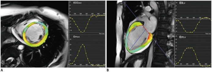
Fig. 1 Myocardial strain measurement by feature tracking method in 43-year-old female patient with acute myocarditis.After endocardial and epicardial borders of LV were traced semi-automatically, software (CVI42) automatically tracked endocardial and epicardial borders across frames during cardiac cycle. EccSAX and ErrSAX measurements (A) were obtained using mid-ventricular level short-axis cine views. EllLV and ErrLax measurements (B) were obtained from 2-chamber long-axis view. EccSAX = LV circumferential strain measured from short-axis cine views, EllLV = LV longitudinal strain measured from long-axis cine views, ErrLax = LV radial strain measured from long-axis cine views, ErrSAX = LV radial strain measured from short-axis cine views, LV = left ventricular
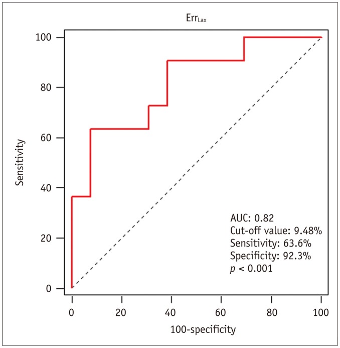
Fig. 2 Receiver operating characteristic curve for prediction of MACE.MACE was defined in terms of cardiac death, heart transplantation, implantable cardioverter defibrillator or pacemaker, rehospitalization following cardiac event, or embolic stroke. AUC = area under the curve, ErrLax = LV radial strain measured from long-axis cine views, LV = left ventricle, MACE = major adverse cardiovascular events
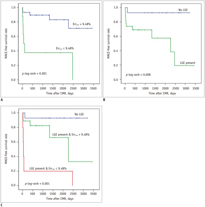
Fig. 3 MACE according to ErrLax or presence of LGE.A. Survivial in patients with ErrLax ≤ 9.48% vs. those with ErrLax > 9.48%. Note that only one patient without LGE experienced MACE dring follow-up. B. Survivial in patients with LGE vs. those without LGE. C. Survivial in patients with ErrLax ≤ 9.48% and presence of LGE vs. those with ErrLax ≤ 9.48% and presence of LGE vs. those without LGE. Patients with LGE and decreased ErrLax (≤ 9.48%) had worse outcome, compared to patients with LGE only. CMR = cardiovascular magnetic resonance, ErrLax = LV radial strain measured from long-axis cine views, LGE = late gadolinium enhancement, LV = left ventricle, MACE = major adverse cardiovascular events
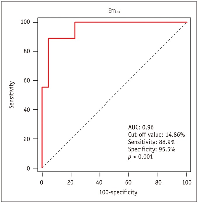
Fig. 4 Receiver operating characteristic curve for prediction of incomplete left ventricular functional recovery.AUC = area under the curve, ErrLax = LV radial strain measured from long-axis cine views, LV = left ventricle
Cited by 6 articles
-
RE: Multi-Parameter CMR Approach in Acute Myocarditis to Improve Diagnosis and Prognostic Stratification
Fausto Pizzino, Antonino Recupero, Pietro Pugliatti, Stefano Maffei, Gianluca Di Bella
Korean J Radiol. 2018;19(2):366-367. doi: 10.3348/kjr.2018.19.2.366.Guidelines for Cardiovascular Magnetic Resonance Imaging from the Korean Society of Cardiovascular Imaging—Part 2: Interpretation of Cine, Flow, and Angiography Data
Jae Wook Lee, Jee Hye Hur, Dong Hyun Yang, Bae Young Lee, Dong Jin Im, Su Jin Hong, Eun Young Kim, Eun-Ah Park, Yeseul Jo, JeongJae Kim, Chul Hwan Park, Hwan Seok Yong
Korean J Radiol. 2019;20(11):1477-1490. doi: 10.3348/kjr.2019.0407.Age of Data in Contemporary Research Articles Published in Representative General Radiology Journals
Ji Hun Kang, Dong Hwan Kim, Seong Ho Park, Jung Hwan Baek
Korean J Radiol. 2018;19(6):1172-1178. doi: 10.3348/kjr.2018.19.6.1172.Guideline for Cardiovascular Magnetic Resonance Imaging from the Korean Society of Cardiovascular Imaging—Part 1: Standardized Protocol
Yeseul Jo, JeongJae Kim, Chul Hwan Park, Jae Wook Lee, Jee Hye Hur, Dong Hyun Yang, Bae Young Lee, Dong Jin Im, Su Jin Hong, Eun Young Kim, Eun-Ah Park, Pan Ki Kim, Hwan Seok Yong
Korean J Radiol. 2019;20(9):1313-1333. doi: 10.3348/kjr.2019.0398.Guidelines for Cardiovascular Magnetic Resonance Imaging from the Korean Society of Cardiovascular Imaging—Part 3: Perfusion, Delayed Enhancement, and T1- and T2 Mapping
Dong Jin Im, Su Jin Hong, Eun-Ah Park, Eun Young Kim, Yeseul Jo, JeongJae Kim, Chul Hwan Park, Hwan Seok Yong, Jae Wook Lee, Jee Hye Hur, Dong Hyun Yang, Bae Young Lee
Korean J Radiol. 2019;20(12):1562-1582. doi: 10.3348/kjr.2019.0411.Guidelines for Cardiovascular Magnetic Resonance Imaging from the Korean Society of Cardiovascular Imaging (KOSCI) - Part 3: Perfusion, Delayed Enhancement, and T1- and T2 Mapping
Dong Jin Im, Su Jin Hong, Eun-Ah Park, Eun Young Kim, Yeseul Jo, Jeong Jae Kim, Chul Hwan Park, Hwan Seok Yong, Jae Wook Lee, Jee Hye Hur, Dong Hyun Yang, Bae-Young Lee
Investig Magn Reson Imaging. 2020;24(1):1-20. doi: 10.13104/imri.2020.24.1.1.
Reference
-
2. Kasper EK, Agema WR, Hutchins GM, Deckers JW, Hare JM, Baughman KL. The causes of dilated cardiomyopathy: a clinicopathologic review of 673 consecutive patients. J Am Coll Cardiol. 1994; 23:586–590. PMID: 8113538.
Article3. Drory Y, Turetz Y, Hiss Y, Lev B, Fisman EZ, Pines A, et al. Sudden unexpected death in persons less than 40 years of age. Am J Cardiol. 1991; 68:1388–1392. PMID: 1951130.4. Mahrholdt H, Wagner A, Deluigi CC, Kispert E, Hager S, Meinhardt G, et al. Presentation, patterns of myocardial damage, and clinical course of viral myocarditis. Circulation. 2006; 114:1581–1590. PMID: 17015795.
Article5. Grün S, Schumm J, Greulich S, Wagner A, Schneider S, Bruder O, et al. Long-term follow-up of biopsy-proven viral myocarditis: predictors of mortality and incomplete recovery. J Am Coll Cardiol. 2012; 59:1604–1615. PMID: 22365425.6. Kindermann I, Kindermann M, Kandolf R, Klingel K, Bültmann B, Müller T, et al. Predictors of outcome in patients with suspected myocarditis. Circulation. 2008; 118:639–648. PMID: 18645053.
Article7. Cooper LT, Baughman KL, Feldman AM, Frustaci A, Jessup M, Kuhl U, et al. The role of endomyocardial biopsy in the management of cardiovascular disease: a scientific statement from the American Heart Association, the American College of Cardiology, and the European Society of Cardiology. Endorsed by the Heart Failure Society of America and the Heart Failure Association of the European Society of Cardiology. J Am Coll Cardiol. 2007; 50:1914–1931. PMID: 17980265.
Article8. Ng AC, Delgado V, Bertini M, Antoni ML, van Bommel RJ, van Rijnsoever EP, et al. Alterations in multidirectional myocardial functions in patients with aortic stenosis and preserved ejection fraction: a two-dimensional speckle tracking analysis. Eur Heart J. 2011; 32:1542–1550. PMID: 21447510.
Article9. Augustine D, Lewandowski AJ, Lazdam M, Rai A, Francis J, Myerson S, et al. Global and regional left ventricular myocardial deformation measures by magnetic resonance feature tracking in healthy volunteers: comparison with tagging and relevance of gender. J Cardiovasc Magn Reson. 2013; 15:8. PMID: 23331550.
Article10. Hsiao JF, Koshino Y, Bonnichsen CR, Yu Y, Miller FA Jr, Pellikka PA, et al. Speckle tracking echocardiography in acute myocarditis. Int J Cardiovasc Imaging. 2013; 29:275–284. PMID: 22736428.
Article11. Morton G, Schuster A, Jogiya R, Kutty S, Beerbaum P, Nagel E. Inter-study reproducibility of cardiovascular magnetic resonance myocardial feature tracking. J Cardiovasc Magn Reson. 2012; 14:43. PMID: 22721175.
Article12. Ibrahim el-SH. Myocardial tagging by cardiovascular magnetic resonance: evolution of techniques--pulse sequences, analysis algorithms, and applications. J Cardiovasc Magn Reson. 2011; 13:36. PMID: 21798021.
Article13. Hor KN, Gottliebson WM, Carson C, Wash E, Cnota J, Fleck R, et al. Comparison of magnetic resonance feature tracking for strain calculation with harmonic phase imaging analysis. JACC Cardiovasc Imaging. 2010; 3:144–151. PMID: 20159640.
Article14. Baeßler B, Schaarschmidt F, Dick A, Michels G, Maintz D. Diagnostic implications of magnetic resonance feature tracking derived myocardial strain parameters in acute myocarditis. Eur J Radiol. 2016; 85:218–227. PMID: 26724669.
Article15. Magnani JW, Dec GW. Myocarditis: current trends in diagnosis and treatment. Circulation. 2006; 113:876–890. PMID: 16476862.16. Friedrich MG, Sechtem U, Schulz-Menger J, Holmvang G, Alakija P, Cooper LT, et al. Cardiovascular magnetic resonance in myocarditis: a JACC white paper. J Am Coll Cardiol. 2009; 53:1475–1487. PMID: 19389557.
Article17. Schultz JC, Hilliard AA, Cooper LT Jr, Rihal CS. Diagnosis and treatment of viral myocarditis. Mayo Clin Proc. 2009; 84:1001–1009. PMID: 19880690.
Article18. Mahrholdt H, Goedecke C, Wagner A, Meinhardt G, Athanasiadis A, Vogelsberg H, et al. Cardiovascular magnetic resonance assessment of human myocarditis: a comparison to histology and molecular pathology. Circulation. 2004; 109:1250–1258. PMID: 14993139.
Article19. Caforio AL, Pankuweit S, Arbustini E, Basso C, Gimeno-Blanes J, Felix SB, et al. Current state of knowledge on aetiology, diagnosis, management, and therapy of myocarditis: a position statement of the European Society of Cardiology working group on myocardial and pericardial diseases. Eur Heart J. 2013; 34:2636–2648. 2648a–2648d. PMID: 23824828.
Article20. Lang RM, Bierig M, Devereux RB, Flachskampf FA, Foster E, Pellikka PA, et al. Recommendations for chamber quantification: a report from the American Society of Echocardiography's Guidelines and Standards Committee and the Chamber Quantification Writing Group, developed in conjunction with the European Association of Echocardiography, a branch of the European Society of Cardiology. J Am Soc Echocardiogr. 2005; 18:1440–1463. PMID: 16376782.
Article21. Sachdeva S, Song X, Dham N, Heath DM, DeBiasi RL. Analysis of clinical parameters and cardiac magnetic resonance imaging as predictors of outcome in pediatric myocarditis. Am J Cardiol. 2015; 115:499–504. PMID: 25554534.
Article22. Buss SJ, Krautz B, Hofmann N, Sander Y, Rust L, Giusca S, et al. Prediction of functional recovery by cardiac magnetic resonance feature tracking imaging in first time ST-elevation myocardial infarction. Comparison to infarct size and transmurality by late gadolinium enhancement. Int J Cardiol. 2015; 183:162–117. PMID: 25675901.
Article23. Onishi T, Saha SK, Ludwig DR, Onishi T, Marek JJ, Cavalcante JL, et al. Feature tracking measurement of dyssynchrony from cardiovascular magnetic resonance cine acquisitions: comparison with echocardiographic speckle tracking. J Cardiovasc Magn Reson. 2013; 15:95. PMID: 24134158.
Article24. André F, Stock FT, Riffel J, Giannitsis E, Steen H, Scharhag J, et al. Incremental value of cardiac deformation analysis in acute myocarditis: a cardiovascular magnetic resonance imaging study. Int J Cardiovasc Imaging. 2016; 32:1093–1101. PMID: 27100527.
Article25. Schuster A, Morton G, Hussain ST, Jogiya R, Kutty S, Asrress KN, et al. The intra-observer reproducibility of cardiovascular magnetic resonance myocardial feature tracking strain assessment is independent of field strength. Eur J Radiol. 2013; 82:296–301. PMID: 23246014.
Article26. Schuster A, Paul M, Bettencourt N, Morton G, Chiribiri A, Ishida M, et al. Cardiovascular magnetic resonance myocardial feature tracking for quantitative viability assessment in ischemic cardiomyopathy. Int J Cardiol. 2013; 166:413–420. PMID: 22130224.
Article27. Schuster A, Stahnke VC, Unterberg-Buchwald C, Kowallick JT, Lamata P, Steinmetz M, et al. Cardiovascular magnetic resonance feature-tracking assessment of myocardial mechanics: Intervendor agreement and considerations regarding reproducibility. Clin Radiol. 2015; 70:989–998. PMID: 26139384.
Article28. Chow LH, Radio SJ, Sears TD, McManus BM. Insensitivity of right ventricular endomyocardial biopsy in the diagnosis of myocarditis. J Am Coll Cardiol. 1989; 14:915–920. PMID: 2794278.
Article29. Abdel-Aty H, Boyé P, Zagrosek A, Wassmuth R, Kumar A, Messroghli D, et al. Diagnostic performance of cardiovascular magnetic resonance in patients with suspected acute myocarditis: comparison of different approaches. J Am Coll Cardiol. 2005; 45:1815–1822. PMID: 15936612.
Article30. Laissy JP, Messin B, Varenne O, Iung B, Karila-Cohen D, Schouman-Claeys E, et al. MRI of acute myocarditis: a comprehensive approach based on various imaging sequences. Chest. 2002; 122:1638–1648. PMID: 12426265.
- Full Text Links
-
- Actions
-
Cited
- CITED
-
- Close
- Share
-
- Similar articles
-
- Cardiovascular Magnetic Resonance Imaging of COVID-19 Myocarditis
- Predictive Value of Cardiac Magnetic Resonance Imaging-Derived Myocardial Strain for Poor Outcomes in Patients with Acute Myocarditis
- Left Atrial Strain Derived From Cardiac Magnetic Resonance Imaging Can Predict Outcomes of Patients With Acute Myocarditis
- Principles and Clinical Applications of Feature-Tracking Cardiac Magnetic Resonance Imaging: A Literature Review
- Cardiac Strain Analysis Using Cine Magnetic Resonance Imaging and Computed Tomography
