Image Reporting and Characterization System for Ultrasound Features of Thyroid Nodules: Multicentric Korean Retrospective Study
- Affiliations
-
- 1Department of Radiology, Severance Hospital, Research Institute of Radiological Science, Yonsei University College of Medicine, Seoul 120-752, Korea.
- 2Department of Biostatistics, Yonsei University College of Medicine, Seoul 102-752, Korea.
- 3Department of Radiology and Research Institute of Radiology, Asan Medical Center, University of Ulsan College of Medicine, Seoul 138-736, Korea.
- 4Department of Radiology, Haeundae Healings Hospital, Busan 612-851, Korea.
- 5Department of Radiology, Konkuk University Medical Center, Konkuk University School of Medicine, Seoul 143-729, Korea.
- 6Department of Radiology, Kangbuk Samsung Hospital, Sungkyunkwan University, Seoul 143-729, Korea.
- 7Department of Radiology, Seoul St. Mary's Hospital, College of Medicine, The Catholic University of Korea, Seoul 137-710, Korea.
- 8Department of Radiology, Gangnam Severance Hospital, Yonsei University College of Medicine, Seoul 135-720, Korea.
- 9Department of Radiology, Seoul National University Hospital, Seoul 110-744, Korea.
- 10Department of Radiology, Thyroid Center, Daerim St. Mary's Hospital, Seoul 150-822, Korea.
- 11Department of Radiology, Dong-A University Medical Center, Busan 602-715, Korea.
- 12Department of Radiology, Hanyang University Hospital, Hanyang University College of Medicine, Seoul 133-791, Korea.
- 13Department of Radiology, Haeundae Paik Hospital, Inje University College of Medicine, Busan 612-862, Korea.
- 14Department of Radiology and Center for Imaging Science, Samsung Medical Center, Sungkyunkwan University School of Medicine, Seoul 135-710, Korea.
- 15Department of Radiology, Human Medical Imaging and Intervention Center, Seoul 137-902, Korea. nndgna@gmail.com
- KMID: 1430053
- DOI: http://doi.org/10.3348/kjr.2013.14.1.110
Abstract
OBJECTIVE
The objective of this retrospective study was to develop and validate a simple diagnostic prediction model by using ultrasound (US) features of thyroid nodules obtained from multicenter retrospective data.
MATERIALS AND METHODS
Patient data were collected from 20 different institutions and the data included 2000 thyroid nodules from 1796 patients. For developing a diagnostic prediction model to estimate the malignant risk of thyroid nodules using suspicious malignant US features, we developed a training model in a subset of 1402 nodules from 1260 patients. Several suspicious malignant US features were evaluated to create the prediction model using a scoring tool. The scores for such US features were estimated by calculating odds ratios, and the risk score of malignancy for each thyroid nodule was defined as the sum of these individual scores. Later, we verified the usefulness of developed scoring system by applying into the remaining 598 nodules from 536 patients.
RESULTS
Among 2000 tumors, 1268 were benign and 732 were malignant. In our multiple regression analysis models, the following US features were statistically significant for malignant nodules when using the training data set: hypoechogenicity, marked hypoechogenicity, non-parallel orientation, microlobulated or spiculated margin, ill-defined margins, and microcalcifications. The malignancy rate was 7.3% in thyroid nodules that did not have suspicious-malignant features on US. Area under the receiver operating characteristic (ROC) curve was 0.867, which shows that the US risk score help predict thyroid malignancy well. In the test data set, the malignancy rates were 6.2% in thyroid nodules without malignant features on US. Area under the ROC curve of the test set was 0.872 when using the prediction model.
CONCLUSION
The predictor model using suspicious malignant US features may be helpful in risk stratification of thyroid nodules.
Keyword
MeSH Terms
Figure
-
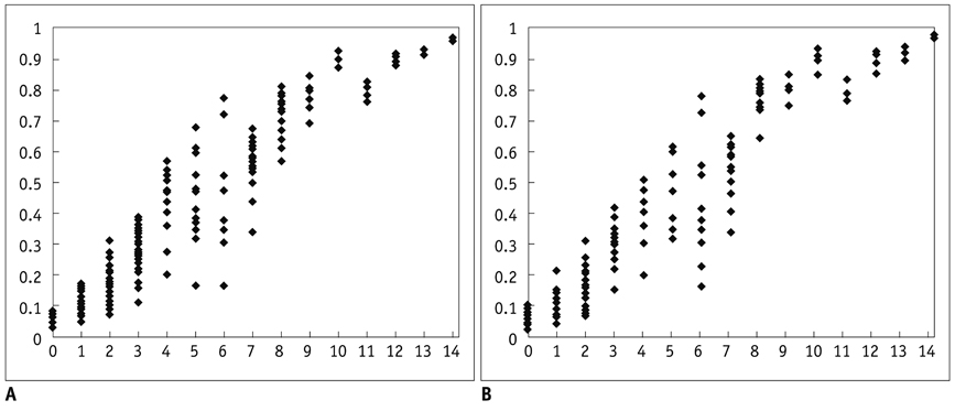
Fig. 1 Scatter plots of predicted probability by total score. Predicted probability of malignancy tended to increase as risk score increased in both data sets. A. Training data set. B. Validation data set.

Fig. 2 Receiver operating characteristic (ROC) curves for training and validation data set based on risk scores from logistic regression model. A. Area under curve is 0.867 at training data set. B. Area under curve is 0.872 at validation data set.
Cited by 4 articles
-
Radiofrequency versus Ethanol Ablation for Treating Predominantly Cystic Thyroid Nodules: A Randomized Clinical Trial
Jung Hwan Baek, Eun Ju Ha, Young Jun Choi, Jin Yong Sung, Jae Kyun Kim, Young Kee Shong
Korean J Radiol. 2015;16(6):1332-1340. doi: 10.3348/kjr.2015.16.6.1332.Complementary Role of Elastography Using Carotid Artery Pulsation in the Ultrasonographic Assessment of Thyroid Nodules: A Prospective Study
Soo Yeon Hahn, Jung Hee Shin, Eun Young Ko, Jung Min Bae, Ji Soo Choi, Ko Woon Park
Korean J Radiol. 2018;19(5):992-999. doi: 10.3348/kjr.2018.19.5.992.Thyroid Imaging Reporting and Data System (TIRADS)
Young Kwak Jin
J Korean Thyroid Assoc. 2013;6(2):106-109. doi: 10.11106/jkta.2013.6.2.106.Ultrasonography Diagnosis of Thyroid Nodules and Cervical Metastatic Lymph Nodes
Dong Gyu Na, Young Hen Lee
Int J Thyroidol. 2016;9(1):1-8. doi: 10.11106/ijt.2016.9.1.1.
Reference
-
1. Park SH, Kim SJ, Kim EK, Kim MJ, Son EJ, Kwak JY. Interobserver agreement in assessing the sonographic and elastographic features of malignant thyroid nodules. AJR Am J Roentgenol. 2009. 193:W416–W423.2. Wienke JR, Chong WK, Fielding JR, Zou KH, Mittelstaedt CA. Sonographic features of benign thyroid nodules: interobserver reliability and overlap with malignancy. J Ultrasound Med. 2003. 22:1027–1031.3. Choi SH, Kim EK, Kwak JY, Kim MJ, Son EJ. Interobserver and intraobserver variations in ultrasound assessment of thyroid nodules. Thyroid. 2010. 20:167–172.4. Lee MJ, Hong SW, Chung WY, Kwak JY, Kim MJ, Kim EK. Cytological results of ultrasound-guided fine-needle aspiration cytology for thyroid nodules: emphasis on correlation with sonographic findings. Yonsei Med J. 2011. 52:838–844.5. Horvath E, Majlis S, Rossi R, Franco C, Niedmann JP, Castro A, et al. An ultrasonogram reporting system for thyroid nodules stratifying cancer risk for clinical management. J Clin Endocrinol Metab. 2009. 94:1748–1751.6. Park JY, Lee HJ, Jang HW, Kim HK, Yi JH, Lee W, et al. A proposal for a thyroid imaging reporting and data system for ultrasound features of thyroid carcinoma. Thyroid. 2009. 19:1257–1264.7. Kwak JY, Han KH, Yoon JH, Moon HJ, Son EJ, Park SH, et al. Thyroid imaging reporting and data system for US features of nodules: a step in establishing better stratification of cancer risk. Radiology. 2011. 260:892–899.8. Hambly NM, Gonen M, Gerst SR, Li D, Jia X, Mironov S, et al. Implementation of evidence-based guidelines for thyroid nodule biopsy: a model for establishment of practice standards. AJR Am J Roentgenol. 2011. 196:655–660.9. American College of Radiology. Breast imaging reporting and data system, breast imaging atlas. 2003. 4th ed. Reston: American College of Radiology.10. Cibas ES, Ali SZ. The Bethesda System for Reporting Thyroid Cytopathology. Thyroid. 2009. 19:1159–1165.11. Moon WJ, Jung SL, Lee JH, Na DG, Baek JH, Lee YH, et al. Benign and malignant thyroid nodules: US differentiation--multicenter retrospective study. Radiology. 2008. 247:762–770.12. American Thyroid Association (ATA) Guidelines Taskforce on Thyroid Nodules and Differentiated Thyroid Cancer. Cooper DS, Doherty GM, Haugen BR, Kloos RT, Lee SL, et al. Revised American Thyroid Association management guidelines for patients with thyroid nodules and differentiated thyroid cancer. Thyroid. 2009. 19:1167–1214.13. Brauer VF, Eder P, Miehle K, Wiesner TD, Hasenclever H, Paschke R. Interobserver variation for ultrasound determination of thyroid nodule volumes. Thyroid. 2005. 15:1169–1175.14. Frates MC, Benson CB, Charboneau JW, Cibas ES, Clark OH, Coleman BG, et al. Management of thyroid nodules detected at US: Society of Radiologists in Ultrasound consensus conference statement. Radiology. 2005. 237:794–800.15. Gharib H, Papini E, Paschke R, Duick DS, Valcavi R, Hegedüs L, et al. American Association of Clinical Endocrinologists, Associazione Medici Endocrinologi, and EuropeanThyroid Association Medical Guidelines for Clinical Practice for the Diagnosis and Management of Thyroid Nodules. Endocr Pract. 2010. 16:Suppl 1. 1–43.16. Kim EK, Park CS, Chung WY, Oh KK, Kim DI, Lee JT, et al. New sonographic criteria for recommending fine-needle aspiration biopsy of nonpalpable solid nodules of the thyroid. AJR Am J Roentgenol. 2002. 178:687–691.17. Papini E, Guglielmi R, Bianchini A, Crescenzi A, Taccogna S, Nardi F, et al. Risk of malignancy in nonpalpable thyroid nodules: predictive value of ultrasound and color-Doppler features. J Clin Endocrinol Metab. 2002. 87:1941–1946.18. Moon WJ, Baek JH, Jung SL, Kim DW, Kim EK, Kim JY, et al. Ultrasonography and the ultrasound-based management of thyroid nodules: consensus statement and recommendations. Korean J Radiol. 2011. 12:1–14.19. Cappelli C, Castellano M, Pirola I, Cumetti D, Agosti B, Gandossi E, et al. The predictive value of ultrasound findings in the management of thyroid nodules. QJM. 2007. 100:29–35.20. Cappelli C, Castellano M, Pirola I, Gandossi E, De Martino E, Cumetti D, et al. Thyroid nodule shape suggests malignancy. Eur J Endocrinol. 2006. 155:27–31.21. Chan BK, Desser TS, McDougall IR, Weigel RJ, Jeffrey RB Jr. Common and uncommon sonographic features of papillary thyroid carcinoma. J Ultrasound Med. 2003. 22:1083–1090.22. Caruso D, Mazzaferri EL. Fine needle aspiration biopsy in the management of thyroid nodules. Endocrinologist. 1991. 1:194–202.23. Kwak JY, Koo H, Youk JH, Kim MJ, Moon HJ, Son EJ, et al. Value of US correlation of a thyroid nodule with initially benign cytologic results. Radiology. 2010. 254:292–300.24. Gharib H, Goellner JR. Fine-needle aspiration biopsy of the thyroid: an appraisal. Ann Intern Med. 1993. 118:282–289.25. Hamburger JI, Hamburger SW. Fine needle biopsy of thyroid nodules: avoiding the pitfalls. N Y State J Med. 1986. 86:241–249.26. Chung WY, Chang HS, Kim EK, Park CS. Ultrasonographic mass screening for thyroid carcinoma: a study in women scheduled to undergo a breast examination. Surg Today. 2001. 31:763–767.27. Moon WJ, Kwag HJ, Na DG. Are there any specific ultrasound findings of nodular hyperplasia ("leave me alone" lesion) to differentiate it from follicular adenoma? Acta Radiol. 2009. 50:383–388.
- Full Text Links
-
- Actions
-
Cited
- CITED
-
- Close
- Share
-
- Similar articles
-
- Thyroid Imaging Reporting and Data System (TIRADS)
- A Glance at the Bethesda System for Reporting Thyroid Cytopathology
- Ultrasound Risk Stratification System of Thyroid Nodule
- Ultrasound Risk Stratification System of Thyroid Nodule
- Korean Thyroid Imaging Reporting and Data System: Current Status, Challenges, and Future Perspectives
