MR Imaging of Solitary Fibrous Tumors in the Head and Neck
- Affiliations
-
- 1Department of Radiology, Daejeon St. Mary's Hospital, College of Medicine, The Catholic University, Korea. rad_1970@yahoo.co.kr
- 2Department of Radiology, Asan Medical Center, University of Ulsan College of Medicine, Korea.
- 3Department of Radiology, Chonnam National University Medical School, Korea.
- 4Department of Radiology, Samsung Medical Center, Sungkyunkwan University School of Medicine, Korea.
- 5Department of Radiology, Ulsan University Hospital, University of Ulsan College of Medicine, Korea.
- 6Department of Diagnostic Pathology, Asan Medical Center, University of Ulsan College of Medicine, Korea.
- KMID: 1102709
- DOI: http://doi.org/10.3348/kjr.2005.6.3.136
Abstract
OBJECTIVE
Solitary fibrous tumor (SFT) is a very rare tumor. The purpose of this study is to determine the MR imaging features of SFT in the intracranial and extracranial head and neck regions. MATERIALS AND METHODS: We retrospectively reviewed six MR images and two CT images of six histologically proven cases of SFT that occurred in four men and two women, and their ages ranged from 46 to 59 years. These imaging findings were correlated with the microscopic findings of their surgical specimens. RESULTS: Six SFTs arose in the meninges (the petrous ridge and the pituitary fossa), the parotid gland, the parapharyngeal space, the buccal space and the maxillary sinus. On the MR images, SFTs in the intracranial and extracranial head and neck regions were mostly isointense to the muscle on the T1-weighted images, they were hyperintense on the T2-weighted images and they all had intense enhancement. On the T1- and T2-weighted images, hypointense lines were observed within in five SFTs. On the CT images, the SFTs were hypodense to the muscle on the unenhanced images and they were heterogeneously enhanced on the contrast-enhanced images. An exceptional case of pituitary SFT was hypointense on the T2-weighted images and it was hyperdense on the unenhanced CT images, which correlated with the increased collagenous component and the cellular compactness. CONCLUSION: The imaging features of SFT are nonspecific; however, SFT should be included in the differential diagnosis of masses involving the intracranial and extracranial head and neck regions.
MeSH Terms
Figure
-
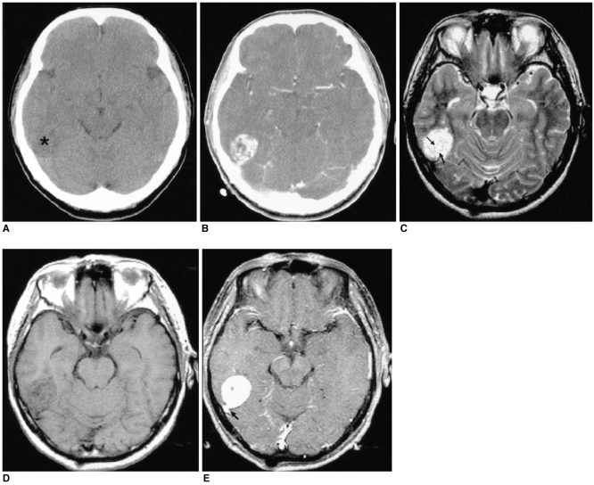
Fig. 1 Images of a 56-year-old man presenting with headache (case 1).A. The axial unenhanced CT image shows an ill-defined low density lesion abutting the temporal lobe (*).B. The axial contrast-enhanced CT image demonstrates the lesion to be a well-defined, heterogeneously well-enhanced, rounded and contoured mass.C. The axial T2-weighted MR image (4000/96 [TR/TE]) shows the lesion to be a well-defined, round mass that is heterogeneously hyperintense to the muscle and there are multiple, hypointense streaks (arrows) within the mass.D. The axial T1-weighted MR image (500/11) shows an isointense mass. The hypointense streaks on the T2-weighted image are also hypointense.E. The axial contrast-enhanced T1-weighted MR images (500/11) reveal homogeneous enhancement of the mass that is based on the petrous ridge and it has a dural 'tail' (arrow). The streaks are well enhanced, as same as the other portions of the mass.
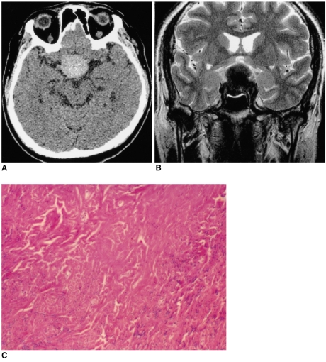
Fig. 2 Images of a 56-year-old man presenting with visual disturbance (case 2).A. The axial unenhanced CT image shows the lesion to be well-defined and hyperdense.B. The T2-weighted axial MR image (4000/98 [TR/TE]) shows a well-defined 'figure eight' shaped mass that is hypointense to the muscle in the pituitary fossa and it has a suprasellar extension.C. Photomicrograph shows the prominent collagenous tissue with cellular areas that are composed of haphazardly arranged spindle cells. (hematoxylin-eosin stain, original A B magnification (100))

Fig. 3 Images of a 59-year-old man presenting with a painless left periauricular mass (case 3).A. The T1-weighted axial MR image shows a well-defined, dumbbell-shaped mass (arrows) that is isointense to the muscle in both the left parapharyngeal space and the parotid space with widening of the stylomandibular tunnel. The mass medially displaces the parapharyngeal fat and the carotid sheath. Multiple hypointense streaks are seen in the mass (open arrow).B. The T2-weighted axial MR image shows the heterogeneous high signal intensity of the mass (arrows). The streaks are also demonstrated as low signal intensity (open arrow).C. The contrast-enhanced T1-weighted axial MR image reveals the homogeneously strong enhancement of the mass. The streaks were less enhanced than in the other portion of the tumor.D. Photomicrograph shows that the tumor is composed of a haphazard proliferation of spindle cells separated from the hyalinized collagen tissue (hematoxylin-eosin, magnification × 200). The inlet shows that the tumor cells and the capillary endothelial cells have immunohistochemically positive CD34 results; this finding is consistent with that of solitary fibrous tumor (original magnification × 200).
Cited by 2 articles
-
A Case of Solitary Fibrous Tumor in the Cheek
Hak Geon Kim, Dong Hoon Kang, Jung Soo Kim, Sung Jae Heo
J Rhinol. 2018;25(1):43-46. doi: 10.18787/jr.2018.25.1.43.Malignant Solitary Fibrous Tumor of the Parotid Gland
Dae Hwan Kim, Ki Ju Cho, Jin Pyeong Kim, Seung Hoon Woo
Korean J Otorhinolaryngol-Head Neck Surg. 2017;60(10):522-526. doi: 10.3342/kjorl-hns.2016.16719.
Reference
-
1. Klemperer P, Rabin CB. Primary neoplasms of the pleura: a report of five cases. Arch Pathol. 1931; 11:385–412.
Article2. England DM, Hochholzer L, McCarthy MJ. Localized benign and malignant tumors of the pleura. A clinicopathologic review of 223 cases. Am J Surg Pathol. 1989; 13:640–658. PMID: 2665534.3. Dervan PA, Tobin B, O'Connor M. Solitary (localized) fibrous mesothelioma: evidence against mesothelial cell origin. Histopathology. 1986; 10:867–875. PMID: 2428726.
Article4. Goodlad JR, Fletcher CD. Solitary fibrous tumor arising at unusual sites: analysis of a series. Histopathology. 1991; 19:515–522. PMID: 1786936.5. Shin JH, Sung IY, Suh JH, Yang SO, Jeong YK, Lee JH, et al. Solitary fibrous tumor in the buccal space: MR Findings with pathologic correlation. AJNR Am J Neuroradiol. 2001; 22:1890–1892. PMID: 11733322.6. Jeong AK, Lee HK, Kim SY, Cho KJ. Solitary fibrous tumor of the parapharyngeal space: MR imaging findings. AJNR Am J Neuroradiol. 2002; 23:473–475. PMID: 11901021.7. Kim TA, Brunberg JA, Pearson JP, Ross DA. Solitary fibrous tumor of the paranasal sinuses: CT and MR appearance. AJNR Am J Neuroradiol. 1996; 17:1767–1772. PMID: 8896635.8. Martin AJ, Fisher C, Igbaseimokumo U, Jarosz JM, Dean AF. Solitary fibrous tumor of the meninges: case series and literature review. J Neurooncol. 2001; 54:57–69. PMID: 11763424.9. Ferretti GR, Chiles C, Cox JE, Choplin RH, Coulomb M. Localized benign fibrous tumors of the pleura: MR appearance. J Comput Assist Tomogr. 1997; 21:115–120. PMID: 9022782.
Article10. Kransdorf MJ, Jelinek JS, Moser RP Jr, Utz JA, Hudson TM, Neal J, et al. Magnetic resonance appearance of fibromatosis. A report of 14 cases and review of the literature. Skeletal Radiol. 1990; 19:495–499. PMID: 2255947.11. Pizzolitto S, Falconieri G, DeMaglio G. Solitary fibrous tumor of the spinal cord: a clinicopathologic study of two cases. Annals of Diagnostic Pathology. 2004; 8:268–275. PMID: 15494932.
Article12. Kessler A, Lapinsky J, Berenholz L, Sarfaty S, Segal A. Solitary fibrous tumor of the nasal cavity. Otolaryngol Head Neck Surg. 1999; 121:826–888. PMID: 10580246.
Article13. Sato J, Asakura K, Yokoyama Y, Satoh M. Solitary fibrous tumor of the parotid gland extending to the parapharyngeal space. Eur Arch Otorhinolaryngol. 1998; 255:18–21. PMID: 9592669.
Article14. Lee KS, Im J-G, Choe KO, Kim CJ, Lee BH. CT findings in benign fibrous mesothelioma of the pleura: pathologic correlation in nine patients. AJR Am J Roentgenol. 1992; 158:983–986. PMID: 1566702.
Article
