Age and Retinal Nerve Fiber Layer Thickness Measured by Spectral Domain Optical Coherence Tomography
- Affiliations
-
- 1Department of Ophthalmology, Gachon University Gil Hospital, Incheon, Korea.
- 2Department of Ophthalmology, Kim's Eye Hospital, Myung-Gok Eye Research Institute, Konyang University College of Medicine, Seoul, Korea. brainh@hanmail.net
- 3Department of Ophthalmology, Armed Forces Capital Hospital, Seongnam, Korea.
- 4Department of Radiation Oncology, Korea University College of Medicine, Seoul, Korea.
- 5Department of Ophthalmology, Korea University College of Medicine, Seoul, Korea.
- KMID: 1376120
- DOI: http://doi.org/10.3341/kjo.2012.26.3.163
Abstract
- PURPOSE
To evaluate the association between age and peripapillary retinal nerve fiber layer (RNFL) thickness measured by Cirrus high-definition (HD) spectral domain optical coherence tomography (OCT) in healthy Korean subjects.
METHODS
A total of 302 eyes from 155 healthy Korean subjects (age range, 20 to 79 years) underwent RNFL thickness measurements using the Cirrus HD-OCT. Average, quadrant, and clock-hour RNFL thickness parameters were analyzed in terms of age using linear mixed effect models.
RESULTS
Average RNFL demonstrated a slope of -2.1 microm per decade of age (p < 0.001). In quadrant analysis, superior (-3.4 microm/decade, p < 0.001) and inferior (-2.9 microm/decade, p < 0.001) quadrants showed steeper slopes, whereas temporal (-1.1 microm/decade, p < 0.001) and nasal (-1.0 microm/decade, p < 0.001) quadrants revealed shallower slopes. Among the 12 clock-hour sectors, clock hours 6 (-4.5 microm/decade, p < 0.001) and 1 (-4.1 microm/decade, p < 0.001) showed the greatest tendency to decline with age; RNFLs of the 3 (-0.2 microm/decade, p = 0.391) and 4 (-0.6 microm/decade, p = 0.052) o'clock hour sectors did not show significant decay.
CONCLUSIONS
RNFL thickness was associated with age, especially in superior and inferior areas. The topographic distribution of correlation between age and RNFL thickness was not uniform.
Keyword
MeSH Terms
Figure
-
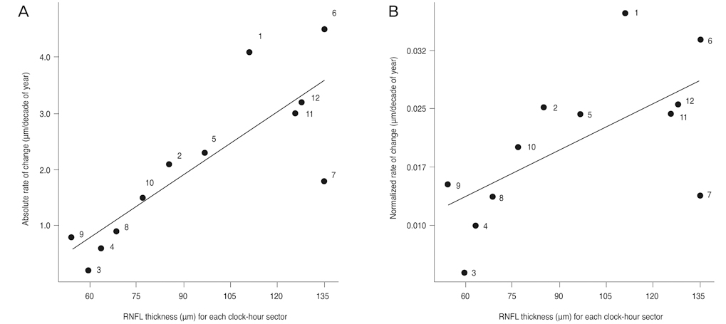
Fig. 1 The relationship between absolute (A), normalized (B) slopes of retinal nerve fiber layer (RNFL) thickness, and RNFL thickness of each clock-hour sector (numbers indicate the clock-hour sectors). Both absolute and normalized rates of RNFL thickness slopes were significantly associated with the average RNFL thickness of each sector (Spearman's rho = 0.853, p < 0.001 for absolute slope; Spearman's rho = 0.661, p = 0.018 for normalized slope).
Cited by 3 articles
-
Retinal Nerve Fiber Layer and Macular Retinal Ganglion Cell Layer Thicknesses in Healthy Korean Children
Yeji Kim, Young Hoon Hwang
J Korean Ophthalmol Soc. 2019;60(9):874-880. doi: 10.3341/jkos.2019.60.9.874.Age-Related Differences of Spectral-Domain Optical Coherence Tomography Data in Koreans
Ji Young Suh, Hong Ryung Seo, Sae Heun Rho
J Korean Ophthalmol Soc. 2013;54(2):289-295. doi: 10.3341/jkos.2013.54.2.289.Retinal Nerve Fiber Layer Thickness Measured by Spectral Domain Optical Coherence Tomography in Healthy Koreans
Youna Choi, Byung Joo Cho
J Korean Ophthalmol Soc. 2018;59(6):537-542. doi: 10.3341/jkos.2018.59.6.537.
Reference
-
1. Schuman JS, Hee MR, Puliafito CA, et al. Quantification of nerve fiber layer thickness in normal and glaucomatous eyes using optical coherence tomography. Arch Ophthalmol. 1995. 113:586–596.2. Lee EJ, Kim TW, Weinreb RN, et al. Trend-based analysis of retin al nerve fiber layer thickness measured by optical coherence tomography in eyes with localized nerve fiber layer defects. Invest Ophthalmol Vis Sci. 2011. 52:1138–1144.3. Leung CK, Liu S, Weinreb RN, et al. Evaluation of retinal nerve fiber layer progression in glaucoma a prospective analysis with neuroretinal rim and visual field progression. Ophthalmology. 2011. 118:1551–1557.4. Dolman CL, McCormick AQ, Drance SM. Aging of the optic nerve. Arch Ophthalmol. 1980. 98:2053–2058.5. Balazsi AG, Rootman J, Drance SM, et al. The effect of age on the nerve fiber population of the human optic nerve. Am J Ophthalmol. 1984. 97:760–766.6. Johnson BM, Miao M, Sadun AA. Age-related decline of human optic nerve axon populations. Age. 1987. 10:5–9.7. Jonas JB, Muller-Bergh JA, Schlotzer-Schrehardt UM, Naumann GO. Histomorphometry of the human optic nerve. Invest Ophthalmol Vis Sci. 1990. 31:736–744.8. Frenkel S, Morgan JE, Blumenthal EZ. Histological measurement of retinal nerve fibre layer thickness. Eye (Lond). 2005. 19:491–498.9. Chi QM, Tomita G, Inazumi K, et al. Evaluation of the effect of aging on the retinal nerve fiber layer thickness using scanning laser polarimetry. J Glaucoma. 1995. 4:406–413.10. Toprak AB, Yilmaz OF. Relation of optic disc topography and age to thickness of retinal nerve fibre layer as measured using scanning laser polarimetry, in normal subjects. Br J Ophthalmol. 2000. 84:473–478.11. Poinoosawmy D, Fontana L, Wu JX, et al. Variation of nerve fibre layer thickness measurements with age and ethnicity by scanning laser polarimetry. Br J Ophthalmol. 1997. 81:350–354.12. Da Pozzo S, Iacono P, Marchesan R, et al. The effect of ageing on retinal nerve fibre layer thickness: an evaluation by scanning laser polarimetry with variable corneal compensation. Acta Ophthalmol Scand. 2006. 84:375–379.13. Schuman JS, Hee MR, Puliafito CA, et al. Quantification of nerve fiber layer thickness in normal and glaucomatous eyes using optical coherence tomography. Arch Ophthalmol. 1995. 113:586–596.14. Varma R, Bazzaz S, Lai M. Optical tomography-measured retinal nerve fiber layer thickness in normal latinos. Invest Ophthalmol Vis Sci. 2003. 44:3369–3373.15. Budenz DL, Anderson DR, Varma R, et al. Determinants of normal retinal nerve fiber layer thickness measured by Stratus OCT. Ophthalmology. 2007. 114:1046–1052.16. Parikh RS, Parikh SR, Sekhar GC, et al. Normal age-related decay of retinal nerve fiber layer thickness. Ophthalmology. 2007. 114:921–926.17. Feuer WJ, Budenz DL, Anderson DR, et al. Topographic differences in the age-related changes in the retinal nerve fiber layer of normal eyes measured by Stratus optical coherence tomography. J Glaucoma. 2011. 20:133–138.18. Sung KR, Wollstein G, Bilonick RA, et al. Effects of age on optical coherence tomography measurements of healthy retinal nerve fiber layer, macula, and optic nerve head. Ophthalmology. 2009. 116:1119–1124.19. Mok KH, Lee VW, So KF. Retinal nerve fiber layer measurement of the Hong Kong Chinese population by optical coherence tomography. J Glaucoma. 2002. 11:481–483.20. Kanamori A, Escano MF, Eno A, et al. Evaluation of the effect of aging on retinal nerve fiber layer thickness measured by optical coherence tomography. Ophthalmologica. 2003. 217:273–278.21. Kanno M, Nagasawa M, Suzuki M, Yamashita H. Peripapillary retinal nerve fiber layer thickness in normal Japanese eyes measured with optical coherence tomography. Jpn J Ophthalmol. 2010. 54:36–42.22. Bendschneider D, Tornow RP, Horn FK, et al. Retinal nerve fiber layer thickness in normals measured by spectral domain OCT. J Glaucoma. 2010. 19:475–482.23. Knight OJ, Chang RT, Feuer WJ, Budenz DL. Comparison of retinal nerve fiber layer measurements using time domain and spectral domain optical coherent tomography. Ophthalmology. 2009. 116:1271–1277.24. Sung KR, Kim DY, Park SB, Kook MS. Comparison of retinal nerve fiber layer thickness measured by Cirrus HD and Stratus optical coherence tomography. Ophthalmology. 2009. 116:1264–1270. 1270.e125. Mikelberg FS, Drance SM, Schulzer M, et al. The normal human optic nerve. Axon count and axon diameter distribution. Ophthalmology. 1989. 96:1325–1328.
- Full Text Links
-
- Actions
-
Cited
- CITED
-
- Close
- Share
-
- Similar articles
-
- Retinal Nerve Fiber Layer Thickness Measured by Spectral Domain Optical Coherence Tomography in Healthy Koreans
- Spectral-Domain Optical Coherence Tomography Findings in Acute Central Retinal Artery Occlusion
- Decreased Retinal Thickness in Patients With Alzheimer's Disease
- Influence of Epiretinal Membranes on the Retinal Nerve Fiber Layer Thickness Measured by Spectral Domain Optical Coherence Tomography in Glaucoma
- Comparison of Retinal Nerve Fiber Layer Thickness Measured by Spectral-Domain and Time-Domain Optical Coherence Tomography
