Partial Trisomy 1q41 Syndrome Delineated by Whole Genomic Array Comparative Genome Hybridization
- Affiliations
-
- 1Department of Rehabilitation Medicine, Pusan National University, School of Medicine, Busan, Korea.
- 2Department of Pediatrics, Pusan National University, School of Medicine, Busan, Korea.
- 3Department of Laboratory Medicine, Pusan National University, School of Medicine, Busan, Korea. mindcatch@hanmail.net
- 4Medical Research Institute, Pusan National University, Busan, Korea.
- 5Department of Laboratory Medicine, Asan Medical Center, University of Ulsan College of Medicine, Seoul, Korea.
- KMID: 1107538
- DOI: http://doi.org/10.3346/jkms.2008.23.6.1097
Abstract
- Partial trisomy 1q syndrome is a rare chromosomal abnormality. We report on a male infant with 46,XY,der(11)t(1;11)(q41;p15.5) due to unbalanced segregation of the maternal reciprocal balanced translocation 46,XX,t(1;11)(q41;p15.5). The baby presented with a mild phenotype, characterized by a triangular face, almond-shaped eyes, low ears, short stature with relatively long legs, and mild psychomotor retardation. We utilized whole genomic array comparative genome hybridization (CGH) with 4,000 selected bacterial artificial chromosomes (BACs) to define the chromosomal breakpoints and to delineate the extent of the partial trisomy in more detail. To our knowledge, this is the first case of nearly pure "partial trisomy 1q41" defined by whole genomic array CGH.
MeSH Terms
Figure
-
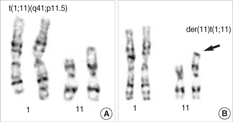
Fig. 1 Karyotype of the proband's mother (A) and the proband (B).

Fig. 2 Ratio plots from array data for chromosomes 1 and 11 of the proband. Each ratio plot is the average of normalized data from two independent arrays. Normalized data from the array in which the test sample was labeled with Cy3 is shown in red while that with Cy5 is shown in blue. (A) Chromosome 1, showing a contiguous duplication on the q-arm. (B) Chromosome 11, showing two-clone deletions of BAC_1341 and BAC_5807 on the p-arm (arrow). The duplicated signals by array CGH in 11q12.2 were considered as copy-number variations because the same signals were also present in the array CGH of proband's mother (arrowhead).
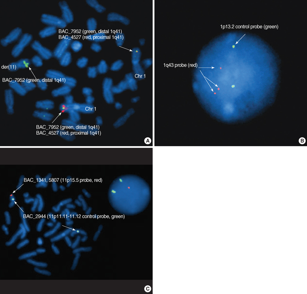
Fig. 3 FISH analysis using BAC probes. (A) BAC_4527 (red; proximal to the interval) and BAC_7952 (green) showing translocation break region of 1q41. (B) 1q43 probe (red, BAC_4069) showed intact signals (a continuous gene rearrangement at 1q41-qter) implicating that no duplication pattern around at 1q43 of the array CGH result was false. (C) BAC_1341 and BAC_5807 (red; distal to the deletion boundary of 11p15.5) were showed a red signal loss of der(11), which confirmed the deletion pattern by the array CGH.
Cited by 2 articles
-
Miller-Dieker Syndrome with der(17)t(12;17)(q24.33;p13.3)pat Presenting with a Potential Risk of Mis-identification as a
de novo Submicroscopic Deletion of 17p13.3
Young Jin Kim, Shin Yun Byun, Seon A Jo, Yong Beom Shin, Eun Hae Cho, Eun Yup Lee, Sang-Hyun Hwang
Korean J Lab Med. 2011;31(1):49-53. doi: 10.3343/kjlm.2011.31.1.49.A Case of Partial Trisomy 2p23-pter Syndrome with Trisomy 18p Due to a
de novo Supernumerary Marker Chromosome
Jong Ho Lee, Hee Soon Cho, Eun Sil Lee, Bo-Chan Jung
Korean J Lab Med. 2010;30(3):312-317. doi: 10.3343/kjlm.2010.30.3.312.
Reference
-
1. Rasmussen SA, Frias JL, Lafer CZ, Eunpu DL, Zackai EH. Partial duplication 1q: report of four patients and review of the literature. Am J Med Genet. 1990. 36:137–143.
Article2. Emberger W, Petek E, Kroisel PM, Zierler H, Wagner K. Clinical and molecular cytogenetic characterization of two patients with partial trisomy 1q41-qter: further delineation of partial trisomy 1q syndrome. Am J Med Genet. 2001. 104:312–318.
Article3. DuPont BR, Huff RW, Ridgway LE, Stratton RF, Moore CM. Prenatal diagnosis of partial trisomy 1q using fluorescent in situ hybridization. Am J Med Genet. 1994. 50:21–27.
Article4. Clark BJ, Lowther GW, Lee WR. Congenital ocular defects associated with an abnormality of the human chromosome 1: trisomy 1q32-qter. J Pediatr Ophthalmol Strabismus. 1994. 31:41–45.
Article5. Duba HC, Erdel M, Loffler J, Bereuther L, Fischer H, Utermann B, Utermann G. Detection of a de novo duplication of 1q32-qter by fluorescence in situ hybridisation in a boy with multiple malformations: further delineation of the trisomy 1q syndrome. J Med Genet. 1997. 34:309–313.
Article6. Chia NL, Bousfield LR, Poon CC, Trudinger BJ. Trisomy (1q)(q42----qter): confirmation of a syndrome. Clin Genet. 1988. 34:224–229.7. Verschuuren-Bemelmans CC, Leegte B, Hodenius TM, Cobben JM. Trisomy 1q42--> qter in a sister and brother: further delineation of the "trisomy 1q42--> qter syndrome". Am J Med Genet. 1995. 58:83–86.8. Concolino D, Cinti R, Ferraro L, Moricca MT, Strisciuglio P. Partial trisomy 1(q42-->qter): a new case with a mild phenotype. J Med Genet. 1998. 35:75–77.9. Pinkel D, Albertson DG. Comparative genomic hybridization. Annu Rev Genomics Hum Genet. 2005. 6:331–354.
Article10. Moon HJ, Yim SV, Lee WK, Jeon YW, Kim YH, Ko YJ, Lee KS, Lee KH, Han SI, Rha HK. Identification of DNA copy-number aberrations by array-comparative genomic hybridization in patients with schizophrenia. Biochem Biophys Res Commun. 2006. 344:531–539.
Article11. Park SJ, Jeong SY, Kim HJ. Y chromosome loss and other genomic alterations in hepatocellular carcinoma cell lines analyzed by CGH and CGH array. Cancer Genet Cytogenet. 2006. 166:56–64.
Article
- Full Text Links
-
- Actions
-
Cited
- CITED
-
- Close
- Share
-
- Similar articles
-
- Partial Trisomy 1q41 Syndrome Delineated by Whole Genomic Array Comparative Genome Hybridization
- Array-based Comparative Genomic Hybridization and Its Application to Cancer Genomes and Human Genetics
- Next generation sequencing and array-based comparative genomic hybridization for molecular diagnosis of pediatric endocrine disorders
- Current Status and Future Clinical Applications of Array.based Comparative Genomic Hybridization
- Low-frequency Mosaicism of Trisomy 14, Missed by Array CGH
