J Korean Ophthalmol Soc.
2014 Oct;55(10):1504-1510. 10.3341/jkos.2014.55.10.1504.
Foveal Shape According to Age and Gender Using Spectral Domain Optical Coherence Tomography
- Affiliations
-
- 1Department of Ophthalmology, Seoul Paik Hospital, Inje University College of Medicine, Seoul, Korea.
- 2Department of Ophthalmology, Sanggye Paik Hospital, Inje University College of Medicine, Seoul, Korea. eyedoctor@freechal.com
- KMID: 2216870
- DOI: http://doi.org/10.3341/jkos.2014.55.10.1504
Abstract
- PURPOSE
To compare foveal shapes in Koreans according to age and gender using spectral domain optical coherence tomography (SD-OCT).
METHODS
This study included 230 eyes of 115 healthy adults. The subjects were divided into three groups: group 1 (20-39 years of age), group 2 (40-59 years of age) and group 3 (60-79 years of age). Using spectralis OCT, we measured central foveal thickness (CFT), regional maximal retinal thickness (MRT), pit diameter and pit depth and compared the differences between the groups.
RESULTS
The MRT of the superior, inferior and nasal sides in group 1 was higher than in groups 2 and 3 (p < 0.05). No significant difference was observed in the MRT of the temporal side. Regarding differences based on age, no significant differences in CFT, pit diameter and pit depth were observed. Regarding differences in gender, the temporal regional MRT of males in group 3 was significantly lower than in group 1 and the pit depth of males in group 1 was significantly higher than in groups 2 and 3. Therefore, differences were observed according to gender.
CONCLUSIONS
In the present study, differences in foveal shape were found according to age and gender which should be considered when foveal diseases are evaluated.
Figure
-
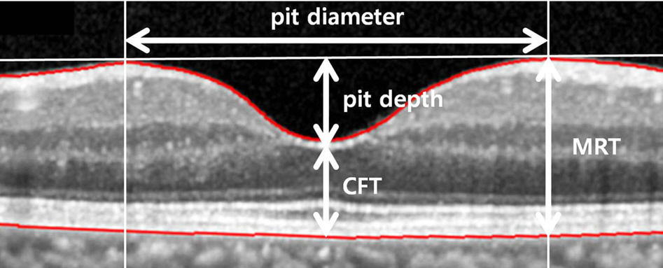
Figure 1. Illustration of the measured parameters. CFT = central foveal thickness; MRT = maximal retinal thickness.
Cited by 1 articles
-
Analysis of Macular Layer Thickness Measured Using Spectral Domain Optical Coherence Tomography in Korean Subjects
Chung Hwan Kim, Sun Young Jin, Young Hoon Lee, Young Suk Chang
J Korean Ophthalmol Soc. 2016;57(2):264-275. doi: 10.3341/jkos.2016.57.2.264.
Reference
-
References
1. Hendrickson A. Organization of the adult primate fovea. Penfold PL, Provis GM, editors. Macular degeneration. Heidelberg, Germany: Springer Verlag;2005. p. 1–23.
Article2. Springer AD, Hendrickson AE. Development of the primate area of high acuity, 3: temporal relationships between pit formation, retinal elongation and cone packing. Vis Neurosci. 2005; 22:171–85.
Article3. van Driel D, Provis JM, Billson FA. Early differentiation of ganglion, amacrine, bipolar, and Muller cells in the developing fovea of human retina. J Comp Neurol. 1990; 291:203–19.
Article4. Provis JM, Diaz CM, Dreher B. Ontogeny of the primate fovea: a central issue in retinal development. Prog Neurobiol. 1998; 54:549–80.5. Ko BW, Shin YW, Lee JM, et al. Comparison of macular thickness measurements between fourier-domain and time-domain optical coherence tomography in normal eyes and eyes with macular diseases. J Korean Ophthalmol Soc. 2009; 50:1661–8.
Article6. Moon SW, Kim ES, Kim YG, et al. The comparison of macular thickness measurements and repeatabilities between time domain and spectral domain OCT. J Korean Ophthalmol Soc. 2009; 50:1050–9.
Article7. Kang NH, Kim HJ, Lee JH. The measurements of macular thickness and volume with SD-OCT in normal eyes. J Korean Ophthalmol Soc. 2011; 52:1182–8.
Article8. Menke MN, Dabov S, Knecht P, Sturm V. Reproducibility of retinal thickness measurements in healthy subjects using spectralis optical coherence tomography. Am J Ophthalmol. 2009; 147:467–72.
Article9. Early Treatment Diabetic Retinopathy Study design and baseline patient characteristics. ETDRS report number 7. Ophthalmology. 1991; 98:741–56.10. Tick S, Rossant F, Ghorbel I, et al. Foveal shape and structure in a normal population. Invest Ophthalmol Vis Sci. 2011; 52:5105–10.
Article11. Kumagai K, Hangai M, Larson E, Ogino N. Foveal thickness in healthy fellow eyes of patients with unilateral macular holes. Am J Ophthalmol. 2013; 156:140–8.
Article12. Song WK, Lee SC, Lee ES, et al. Macular thickness variations with sex, age, and axial length in healthy subjects: a spectral domain-optical coherence tomography study. Invest Ophthalmol Vis Sci. 2010; 51:3913–8.
Article13. Zou H, Zhang X, Xu X, Yu S. Quantitative in vivo retinal thickness measurement in chinese healthy subjects with retinal thickness analyzer. Invest Ophthalmol Vis Sci. 2006; 47:341–7.
Article14. Kanai K, Abe T, Murayama K, Yoneya S. [Retinal thickness and changes with age]. Nihon Ganka Gakkai Zasshi. 2002; 106:162–5.
Article15. Neuville JM, Bronson-Castain K, Bearse MA Jr, et al. OCT reveals regional differences in macular thickness with age. Optom Vis Sci. 2009; 86:E810–6.
Article16. Guedes V, Schuman JS, Hertzmark E, et al. Optical coherence tomography measurement of macular and nerve fiber layer thickness in normal and glaucomatous human eyes. Ophthalmology. 2003; 110:177–89.
Article17. Kang JH, Kim SA, Song WG, Yoon HS. Macular thickness changes with age in normal subjects measured by optical coherence tomography. J Korean Ophthalmol Soc. 2004; 45:592–8.18. Kim SH, Choi KS, Lee SJ. Macular thickness changes with age and gender in emmetropia using spectral domain optical coherence tomography. J Korean Ophthalmol Soc. 2011; 52:299–307.
Article19. Lee YJ. Analysis of factors associated with variability in measures obtained by spectral domain optical coherence tomography. J Korean Ophthalmol Soc. 2012; 53:639–46.
Article20. Wagner-Schuman M, Dubis AM, Nordgren RN, et al. Race- and sex-related differences in retinal thickness and foveal pit morphology. Invest Ophthalmol Vis Sci. 2011; 52:625–34.
Article21. Ooto S, Hangai M, Sakamoto A, et al. Three-dimensional profile of macular retinal thickness in normal Japanese eyes. Invest Ophthalmol Vis Sci. 2010; 51:465–73.
Article22. Kashani AH, Zimmer-Galler IE, Shah SM, et al. Retinal thickness analysis by race, gender, and age using Stratus OCT. Am J Ophthalmol. 2010; 149:496–502.e1.
Article23. Lim MC, Hoh ST, Foster PJ, et al. Use of optical coherence tomography to assess variations in macular retinal thickness in myopia. Invest Ophthalmol Vis Sci. 2005; 46:974–8.
Article24. Kiernan DF, Hariprasad SM, Chin EK, et al. Prospective comparison of cirrus and stratus optical coherence tomography for quantifying retinal thickness. Am J Ophthalmol. 2009; 147:267–75.e2.
Article25. Yu X, Tang Y, Li F, et al. Protection against hydrogen peroxide-induced cell death in cultured human retinal pigment epithelial cells by 17beta-estradiol: a differential gene expression profile. Mech Ageing Dev. 2005; 126:1135–45.26. Kim SH, Park JY, Park TK, Ohn YH. Use of spectral-domain optical coherence tomography to analyze macular thickness according to refractive error. J Korean Ophthalmol Soc. 2011; 52:1286–95.
Article27. Lim MC, Hoh ST, Foster PJ, et al. Use of optical coherence tomography to assess variations in macular retinal thickness in myopia. Invest Ophthalmol Vis Sci. 2005; 46:974–8.
Article
- Full Text Links
-
- Actions
-
Cited
- CITED
-
- Close
- Share
-
- Similar articles
-
- Foveal Shape According to Age and Gender Using Spectral Domain Optical Coherence Tomography
- A Study of Foveal Shape in Emmetropia and Myopia Using Spectral Domain Optical Coherence Tomography
- Macular Thickness Changes with Age and Gender in Emmetropia Using Spectral Domain Optical Coherence Tomography
- Comparison of Spectral-Domain and Time-Domain Optical Coherence Tomography in Solar Retinopathy
- Choroidal Thickness at the Outside of Fovea in Diabetic Retinopathy Using Spectral-Domain Optical Coherence Tomography
