Retinal Nerve Fiber Layer Defect Associated with Astrocytic Hamartoma in a Patient with Tuberous Sclerosis
- Affiliations
-
- 1The Institute of Vision Research, Department of Ophthalmology, Yonsei University College of Medicine, Seoul, Korea. kcyeye@yuhs.ac
Abstract
- PURPOSE
To report the progression of an astrocytic hamartoma of the right optic nerve head as well as the retina, and the progression of retinal nerve fiber defect associated with astrocytic hamartoma in a patient with tuberous sclerosis.
CASE SUMMARY
A 6-year-old boy with tuberous sclerosis and an astrocytic hamartoma of the right optic nerve head, which was found at the time of ophthalmologie examinations, was referred from the pediatric neurologist for evaluation of the vigabatrin-associated visual field changes. Fundus examination revealed 1/2 disc diameter (DD)-sized astrocytic hamartoma located at the margin of the superior part of the optic nerve. The retina of the left eye was normal. Eighteen months after the first visit, enlarged optic disc hamartoma of the right eye and newly onset retinal astrocytic hamartoma located approximately 1.5 DD inferior to the fovea of the left eye were found. Three years later, an increase in the size of the astrocytic hamartoma of the right optic nerve and development of retinal nerve fiber defects were observed.
CONCLUSIONS
Astrocytic hamartoma in patients with tuberous sclerosis is usually stable without progression. However, in our patient, astrocytic hamartoma showed progression, and development of retinal nerve fiber defects occurred. Regular follow-up is necessary for astrocytic hamartoma in patients with tuberous sclerosis.
MeSH Terms
Figure
-
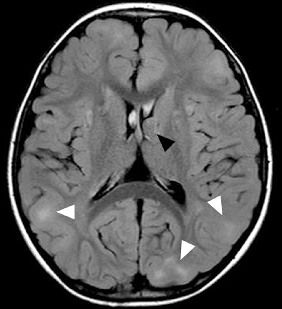
Figure 1. Axial T2-weighted images of MR show that subependymal nodule (black arrow head) projecting into lumen of left lateral ventricle and multiple subtle bilateral cortical and subcortical tubers (white arrow heads).

Figure 2. Development of nerve fiber layer defect associated with astrocytic hamartoma in a patient with tuberous sclerosis. (A) Fundus photograph of the initial visit shows a 1/2 disc diameter-sized “mulberry-like” protruding tu-mor above the right optic disc and normal fellow eye. (B) One year and 6 months after the first visit, fundus photo-graph shows enlarged right optic disc hamartoma. Newly onset retinal astrocytic hamartoma (black arrow) is found on the fundus of the left eye. (C) Three years later, fundus photograph shows development of retinal nerve fiber layer defects (white arrows).
Reference
-
References
1. Curatolo P, Bombardieri R, Jozwiak S. Tuberous sclerosis. Lancet. 2008; 372:657–68.
Article2. Osborne JP, Fryer A, Webb D. Epidemiology of tuberous sclerosis. Ann N Y Acad Sci. 1991; 615:125–7.
Article3. van Slegtenhorst M, de Hoogt R, Hermans C. . Identification of the tuberous sclerosis gene TSC1 on chromosome 9q34. Science. 1997; 277:805–8.4. European Chromosome 16 Tuberous Sclerosis Consortium. Identification and characterization of the tuberous sclerosis gene on chromosome 16 Cell. 1993; 75:1305–15.5. Vogt H. Zur diagnostik der tuberosen sclerose. Z Erforsch Benhandl Fugendl Schwachsinns. 1908; 2:1–16.6. Roach ES, Gomez MR, Northrup H. Tuberous sclerosis complex consensus conference: revised clinical diagnostic criteria. J Child Neurol. 1998; 13:624–8.
Article7. Rowley SA, O'Callaghan FJ, Osborne JP. Ophthalmic manifes-tations of tuberous sclerosis: a population based study. Br J Ophthalmol. 2001; 85:420–3.
Article8. Kiribuchi K, Uchida Y, Fukuyama Y, Maruyama H. High incidence of fundus hamartomas and clinical significance of a fundus score in tuberous sclerosis. Brain Dev. 1986; 8:509–17.
Article9. Töteberg-Harms M, Sturm V, Sel S. . Retinal astrocytomas: long-term follow-up. Klin Monbl Augenheilkd. 2011; 228:337–9.
Article10. Gass JDM. Stereoscopic atlas of macular disease. Diagnosis and treatment. 1st ed.St. Louis: Mosby;1997; 836–43.11. Lagos JC, Gomez MR. Tuberous sclerosis: Reappraisal of a clin-ical entity. Mayo Clin Proc. 1967; 42:26–49.12. Nyboer JH, Robertson DM, Gomez MR. Retinal lesions in tuber-ous sclerosis. Arch Ophthal. 1976; 94:1277–80.
Article13. Yoon DK. A case of tuberous sclerosis. J Korean Ophthalmol Soc. 1974; 15:68–71.14. Kim JH, Shin HH. Two cases of tuberous sclerosis. J Korean Ophthalmol Soc. 1982; 23:477–81.15. Kwon OW, Kim YW. A case of tuberous sclerosis. J Korean Ophthalmol Soc. 1983; 24:645–9.16. Yoo KH, Oum BS. Two cases of tuberous sclerosis. J Korean Ophthalmol Soc. 1987; 28:877–83.17. Kim SM, Hahn YH, Kim SD. Two cases of tuberous sclerosis. J Korean Ophthalmol Soc. 1988; 29:1141–5.18. Ku YJ, Kim EA, Han YB. A case of tuberous sclerosis. J Korean Ophthalmol Soc. 1995; 36:355–60.19. Ji JY, Lee JH, Choi WS. A case of bilateral tuberous sclerosis. J Korean Ophthalmol Soc. 1996; 37:203–9.20. Lee JH, Han KS, Han YB. One case of tuberous sclerosis. J Korean Ophthalmol Soc. 1999; 40:2337–42.21. La TY, Kim CW, Lee YS, Kim MH. A case of retinal astrocytic ha-martoma causing blindness in tuberous sclerosis. J Korean Ophthalmol Soc. 1999; 40:1421–6.22. Dichter MA, Brodie MJ. New antiepileptic drugs. N Engl J Med. 1996; 334:1583–90.
Article23. Ben-Menachem E. Vigabatrin. Epilepsia. 1995; 36:Suppl 2. S95–104.
Article24. Maguire MJ, Hemming K, Wild JM. . Prevalence of visual field loss following exposure to vigabatrin therapy: a systematic review. Epilepsia. 2010; 51:2423–31.
Article25. Roubertie A, Bellet H, Echenne B. Vigabatrin-associated retinal cone system dysfunction. Neurology. 1998; 51:1779–81.
Article26. Miller NR, Johnson MA, Paul SR. . Visual dysfunction in patients receiving vigabatrin: clinical and electrophysiologic findings. Neurology. 1999; 53:2082–7.
Article27. Lawthom C, Smith PE, Wild JM. Nasal retinal nerve fiber layer at-tenuation: a biomarker for vigabatrin toxicity. Ophthalmology. 2009; 116:565–71.
Article28. Clayton LM, Devile M, Punte T. . Patterns of peripapillary reti-nal nerve fiber layer thinning in vigabatrin-exposed individuals. Ophthalmology. 2012; 119:2152–60.
Article29. Roh S, Noecker RJ, Schuman JS. . Effect of optic nerve head drusen on nerve fiber layer thickness. Ophthalmology. 1998; 105:878–85.30. Savino PJ, Glaser JS, Rosenberg MA. A clinical analysis of pseudopapilledema. II. Visual field defects. Arch Ophthalmol. 1979; 97:71–5.
Article31. Sarkies NJ, Sanders MD. Optic disc drusen and episodic visual loss. Br J Ophthalmol. 1987; 71:537–9.
Article32. Shields CL, Benevides R, Materin MA, Shields JA. Optical coher-ence tomography of retinal astrocytic hamartoma in 15 cases. Ophthalmology. 2006; 113:1553–7.
Article
- Full Text Links
-
- Actions
-
Cited
- CITED
-
- Close
- Share
-
- Similar articles
-
- Retinal Nerve Fiber Layer Defect Associated with Astrocytic Hamartoma in a Patient with Tuberous Sclerosis
- Fundus Autofluorescence, Fluorescein Angiography and Spectral Domain Optical Coherence Tomography Findings of Retinal Astrocytic Hamartomas in Tuberous Sclerosis
- A Case of Tuberous Sclerosis
- Indocyanine green angiography of retinal astrocytomas associated with tuberous sclerosis
- A Case of Retinal Astrocytic Hamartoma Causing Blindness in Tuberous Sclerosis
