Wyburn-Mason Syndrome: A Case Report
- Affiliations
-
- 1Department of Ophthalmology, Eulji University Hospital, College of Medicine, Daejeon, Korea. snlee@eul.ji.ac.kr
Abstract
- PURPOSE
To describe a case of Wyburn-Mason Syndrome, which is characterized by arteriovenous malformations in the central nervous system and the retina.
CASE SUMMARY
A 13-year-old girl who underwent neurosurgical intervention for intraventricular hemorrhage was referred to our ophthalmic clinic because brain angiogram and MRI finding revealed arteriovenous malformations (AVMs) that extended from the orbit and optic chiasm along the optic pathway. Fundus and fluorescein angiography of the left eye showed marked dilation and tortuosity of the retinal vessels. Fluorescein angiographic findings showed rapid filling of all vessels and no dye leakage.
CONCLUSIONS
Retinal racemose hemangioma should be considered for Wyburn-Mason syndrome, and the finding of retinal AVMs should warrant cerebral imaging studies including MRI and angiography.
MeSH Terms
Figure
-

Figure 1. Angiograms of the left internal carotid artery (A: Anteroposterior view, B: Right antero-oblique view, C & D: Lateral view). There are AVMs that extends from the orbit and optic chiasm along the optic pathway to the midbrain (o: Ophthalmic artery, i: Internal carotid artery, AVM: AV malformations)
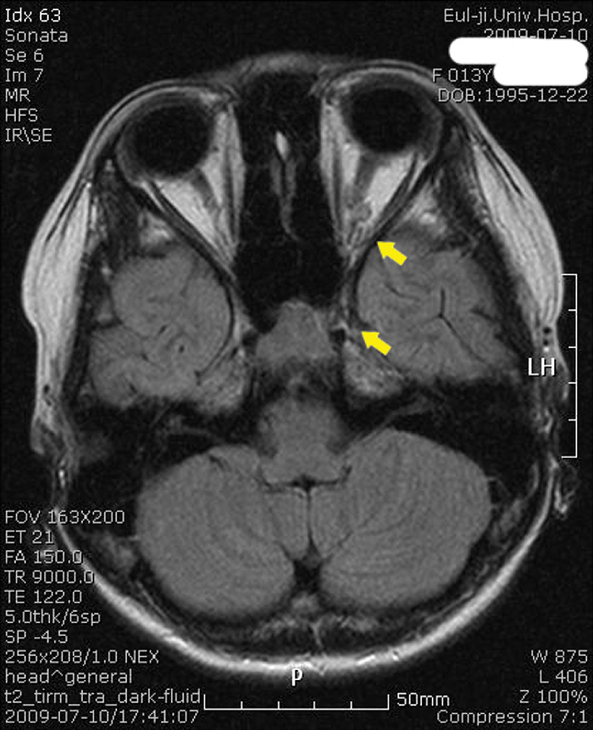
Figure 2. Magnetic resonance imaging shows aggregated enhancing structures (yellow arrow) in the left central orbital space and suprasellar cistern, and around the supraclinoid internal carotid artery.
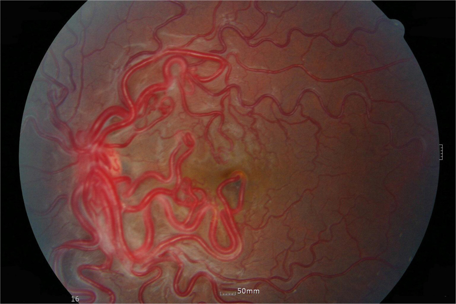
Figure 3. Color fundus photograph of the left eye shows marked dilation and tortuosity of the retinal vessels, “Bag of Worms”

Figure 4. Fluorescein angiograms of the left eye. In the arterial phase, Rapid arterial filling and immediately emptying of fluorescein dye into the vein through the various A-V communications are seen. In the late phase, No leakage is evident.
Reference
-
References
1. Wyburn-Mason R. Arteriovenous aneurysm of midbrain and retina, facial naevi and mental changes. Brain. 1943; 66:163–203.2. Bae HD, Kim BH, Shin DM, et al. A Case of Congenital aberrations Communication of the Retina. J Korean Ophthalmol Soc. 1981; 22:647–51.3. Jang HS, Lee TK, Kim JW. A Case of Retinal Racemose aberrations. J Korean Ophthalmol Soc. 2001; 42:1232–5.4. Bonnet P, Dechaume J, Blanc M. L'anevrisme cirsoide de la retine(Anevrysme racemeux). J de med de Lyon. 1937; 18:165–78.5. Theron J, Newton TH, Hoyt WF. Unilateral retinocephalic vascular malformations. Neuroradiology. 1974; 7:185–96.
Article6. Bech K, Jensen OA. On the frequency of coexisting racemose aberrations of the retina and brain. Acta Psychiatr Neurol Scand. 1961; 36:47–56.7. Goh D, Malik N N, Gilvarry A. Retinal racemose haemangioma directly communicating with a intramuscular facial cavernous haemangioma Br J Ophthalmol. 2004; 88:840–2.8. Magnus H. Aneurysma arteriosovenosum retinale. Virchows Arch Pathol Anat Physiol. 1874; 60:38–43.
Article9. Carmeron ME, Greer CH. Congenital arteriovenous aneurysm of the retina, A post-mortem report. Brit Histol Ophthalmol. 1968; 52:768–72.10. Noden DM. Cell movements and control of patterned tissue assembly during craniofacial development. J Craniofac Genet Dev Biol. 1991; 11:192–213.11. Morgan MK, Johnston IH, De Silva M. Treatment of ophthalmofacial-hypothalamic arteriovenous malformation (Bonnet-Dechaume-Blanc syndrome). Case report. J Neurosurg. 1985; 63:794–6.12. Reck SD, Zacks DN, Eibschitz-Tsimhoni M. Retinal and aberrations arteriovenous malformations: Wyburn-Mason syndrome. J Neuroophthalmol. 2005; 25:205–8.13. Lee AW, Chen CS, Gailloud P, et al. Wyburn-Mason Syndrome Associated with Thyroid Arteriovenous Malformation: A First Case Report. Am J Neuroradiol. 2007; 28:1153–4.
Article
- Full Text Links
-
- Actions
-
Cited
- CITED
-
- Close
- Share
-
- Similar articles
-
- Wyburn-Mason Syndrome: A Case Report
- A Case of Wyburn-Mason Syndrome with Facial Nevus Flammeus and the Associated Optical Coherence Tomography Findings
- A Case of Dyke-Davidoff-Mason Syndrome
- Modified Mason-Allen Suture Bridge Technique: A New Suture Bridge Technique with Improved Tissue Holding by the Modified Mason-Allen Stitch
- Feasibility of York-Mason Operation for Selective Advanced Rectal Cancer
