Clinical Analysis of Acute Radiculopathy after Osteoporotic Lumbar Compression Fracture
- Affiliations
-
- 1Department of Neurosurgery, College of Medicine, Chosun University, Gwangju, Korea. chosunns@chosun.ac.kr
- 2Department of Neurosurgery, Heori Sarang Hospital, Daejeon, Korea.
- 3Department of Internal Medicine, Soonchunhyang University College of Medicine, Seoul, Korea.
- KMID: 2067088
- DOI: http://doi.org/10.3340/jkns.2015.57.1.32
Abstract
OBJECTIVE
The purpose of this study was to analyze the relationship between fracture pattern and the development of acute radiculopathy after osteoporotic lumbar compression fracture.
METHODS
This study included 59 patients who underwent bone cement augmentation for osteoporotic compression fracture below the L2 level, which can lead to radiculopathic radiating pain. The patients were divided into two groups according to the presence of radiculopathy (group A : back pain only; group B : back pain with newly developed radiating pain). We categorized compression fractures into three types by the position of the fracture line. The incidence of newly developed radiculopathy was examined retrospectively for each compression fracture type.
RESULTS
The overall incidence of newly developed leg pain (group B) was 25%, and the frequency increased with descending spinal levels (L2 : 0%, L3 : 22%, L4 : 43%, and L5 : 63%). The back pain-only group (group A) had mostly superior-type fractures. On the other hand, the back pain with radiculopathy group (group B) had mostly inferior-type fractures. Most patients in group B showed significant relief of leg pain as well as back pain after bone cement augmentation.
CONCLUSION
The incidence of a newly developed, radiating pain after osteoporotic compression fractures increased gradually from the L3 to L5 levels. Most of these fractures were of the inferior type, and the bone cement augmentation procedures seemed to be sufficient for relief of both back and radiating pain.
Keyword
MeSH Terms
Figure
-
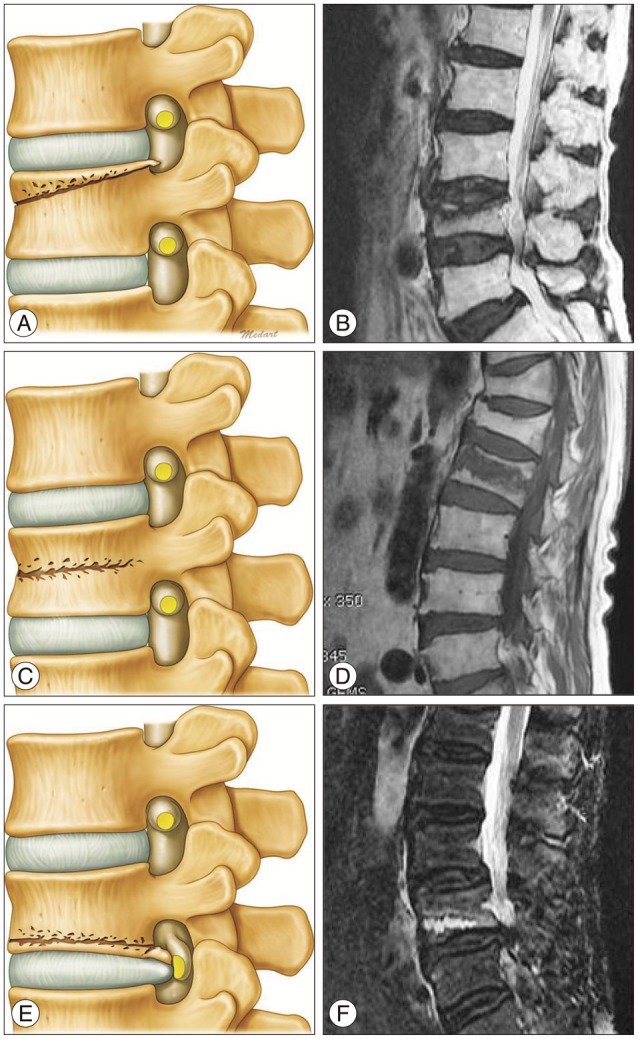
Fig. 1 Three fracture types according to the main position of the fracture line. A and B : Superior type. C and D : Middle type. E and F : Inferior type.
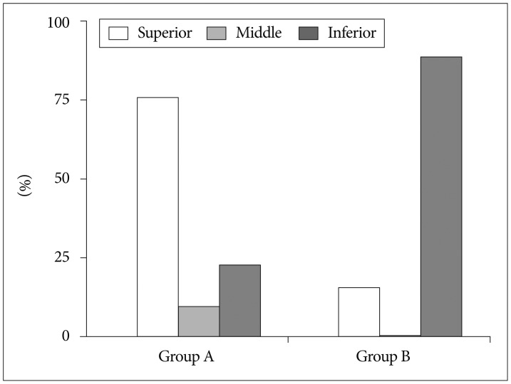
Fig. 2 The intergroup difference in fracture pattern.
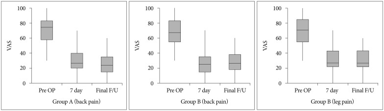
Fig. 3 The improvement in pain score. VAS : visual analogue scale, OP : operation, F/U : follow-up.
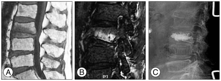
Fig. 4 A 67-year-old woman with back pain and left leg radiating pain (inferior fracture type). A and B : T1-weighted sagittal and fat-suppression images showing osteoporotic compression fracture at L3, and root compression at the intervertebral foramen. C : Simple radiographic image obtained after the vertebroplasty at L3.
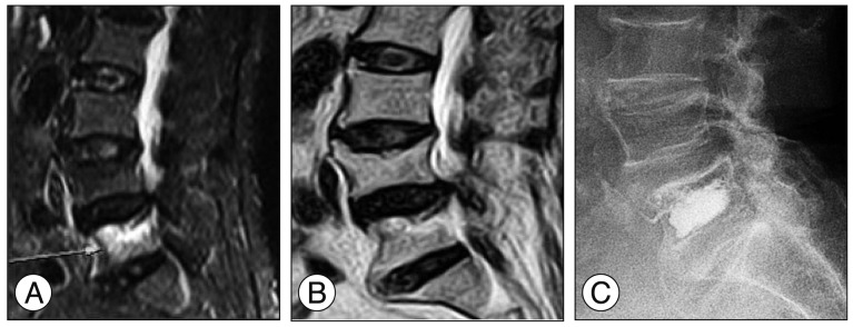
Fig. 5 A-69-year-old woman with back pain and right leg radiating pain (superior fracture type). A and B : T2-weighted and fat-suppression images showing osteoporotic compression fracture at L5 and stenosis at levels L4 and L5 (arrow). C : Simple radiographic image obtained after the vertebroplasty at L5.
Cited by 1 articles
-
Unilateral Biportal Endoscopy as a Treatment for Acute Radiculopathy after Osteoporotic Lumbar Compression Fracture - A Case Report -
Hyoung Bok Kim, Hoon-Jae Chung
J Korean Soc Spine Surg. 2019;26(1):21-25. doi: 10.4184/jkss.2019.26.1.21.
Reference
-
1. Buchbinder R, Osborne RH, Ebeling PR, Wark JD, Mitchell P, Wriedt C, et al. A randomized trial of vertebroplasty for painful osteoporotic vertebral fractures. N Engl J Med. 2009; 361:557–568. PMID: 19657121.
Article2. Chung SK, Lee SH, Kim DY, Lee HY. Treatment of lower lumbar radiculopathy caused by osteoporotic compression fracture : the role of vertebroplasty. J Spinal Disord Tech. 2002; 15:461–468. PMID: 12468971.
Article3. Doo TH, Shin DA, Kim HI, Shin DG, Kim HJ, Chung JH, et al. Clinical relevance of pain patterns in osteoporotic vertebral compression fractures. J Korean Med Sci. 2008; 23:1005–1010. PMID: 19119444.
Article4. Dublin AB, Hartman J, Latchaw RE, Hald JK, Reid MH. The vertebral body fracture in osteoporosis : restoration of height using percutaneous vertebroplasty. AJNR Am J Neuroradiol. 2005; 26:489–492. PMID: 15760853.5. Farrokhi MR, Alibai E, Maghami Z. Randomized controlled trial of percutaneous vertebroplasty versus optimal medical management for the relief of pain and disability in acute osteoporotic vertebral compression fractures. J Neurosurg Spine. 2011; 14:561–569. PMID: 21375382.
Article6. Kanchiku T, Taguchi T, Kawai S. Magnetic resonance imaging diagnosis and new classification of the osteoporotic vertebral fracture. J Orthop Sci. 2003; 8:463–466. PMID: 12898295.
Article7. Katz JN, Harris MB. Clinical practice. Lumbar spinal stenosis. N Engl J Med. 2008; 358:818–825. PMID: 18287604.8. Lee JH, Kwon JT, Kim YB, Suk JS. Segmental deformity correction after balloon kyphoplasty in the osteoporotic vertebral compression fracture. J Korean Neurosurg Soc. 2007; 42:371–376. PMID: 19096572.
Article9. Miller JD, Nader R. Treatment of combined osteoporotic compression fractures and spinal stenosis : use of vertebral augumentation and interspinous process spacer. Spine (Phila Pa 1976). 2008; 33:E717–E720. PMID: 18758354.10. Rao RD, Singrakhia MD. Painful osteoporotic vertebral fracture. Pathogenesis, evaluation, and roles of vertebroplasty and kyphoplasty in its management. J Bone Joint Surg Am. 2003; 85-A:2010–2022. PMID: 14563813.
- Full Text Links
-
- Actions
-
Cited
- CITED
-
- Close
- Share
-
- Similar articles
-
- Unilateral Biportal Endoscopy as a Treatment for Acute Radiculopathy after Osteoporotic Lumbar Compression Fracture: A Case Report
- Clinical Analysis of Acute Radiculopathy after Osteoporotic Lumbar Compression Fracture
- Analysis of Osteoporotic Spinal Compression Fractures in Whole Spine Sagittal MR Images
- Lumbar Nerve Root Compression due to Leakage of Bone Cement after Vertebroplasty
- Measurement of Bone Mineral Density of Lumbar Spine in Osteoporotic Patients Treated with Percutaneous Vertebroplasty
