Progressive Multifocal Leukoencephalopathy in a Patient with Systemic Lupus Erythematosus
- Affiliations
-
- 1Department of Internal Medicine, The Catholic University of Korea College of Medicine, Seoul, Korea.
- 2Department of Internal Medicine, Holy Family Hospital, Bucheon, Korea. rmin6403@hanmail.net
- 3Department of Neurology, The Catholic University of Korea College of Medicine, Seoul, Korea.
- 4Department of Radiology, The Catholic University of Korea College of Medicine, Seoul, Korea.
- KMID: 1270233
- DOI: http://doi.org/10.4078/jkra.2008.15.2.159
Abstract
- Progressive multifocal leukoencephalopathy (PML) is a rare, serious, and usually fatal demyelinating disease that occurs predominantly in severely immunosuppressed patients. The disease is caused by the infection of oligodendrocytes with JC virus that is widely distributed as a latent infection in the general populations. PML has been described mainly in patients infected with the human immunodeficiency virus. However, other immune-suppressed patients including malignancies and organ transplants can be affected with JC virus infection. Recently it is suggested that rheumatologic diseases, including systemic lupus erythematosus (SLE), rheumatoid arthritis, dermatomyositis, polymyositis, wegener`s granulomatosis be known to be at risk of developing PML. We report a case of PML in a patient with SLE.
MeSH Terms
Figure
-
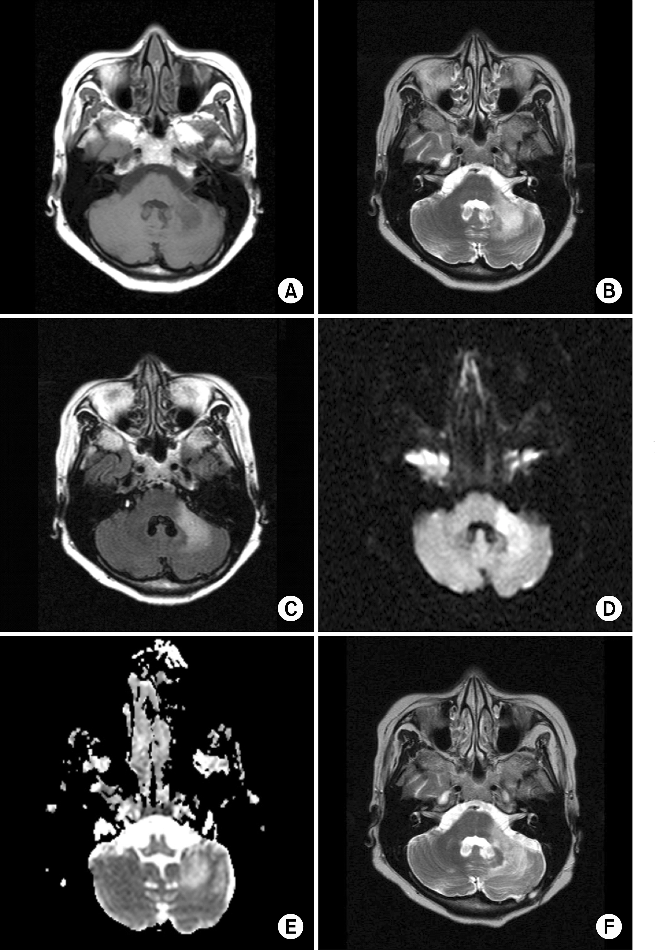
Fig. 1. MR images demonstrate patchy low signal intensity on T1-weighted image (A) and high signal intensity on T2-weighted image (B) in left middle cerebellar peduncle. FLAIR image (C) and diffusion image (D) showing lesions with increased signal affecting left middle cerebellar peduncle. ADC map (E) showing high value in corresponding lesion seen on the FLAIR image. Follw-up T2-weighted image (F) demonstrating further extension of the previous lesions.
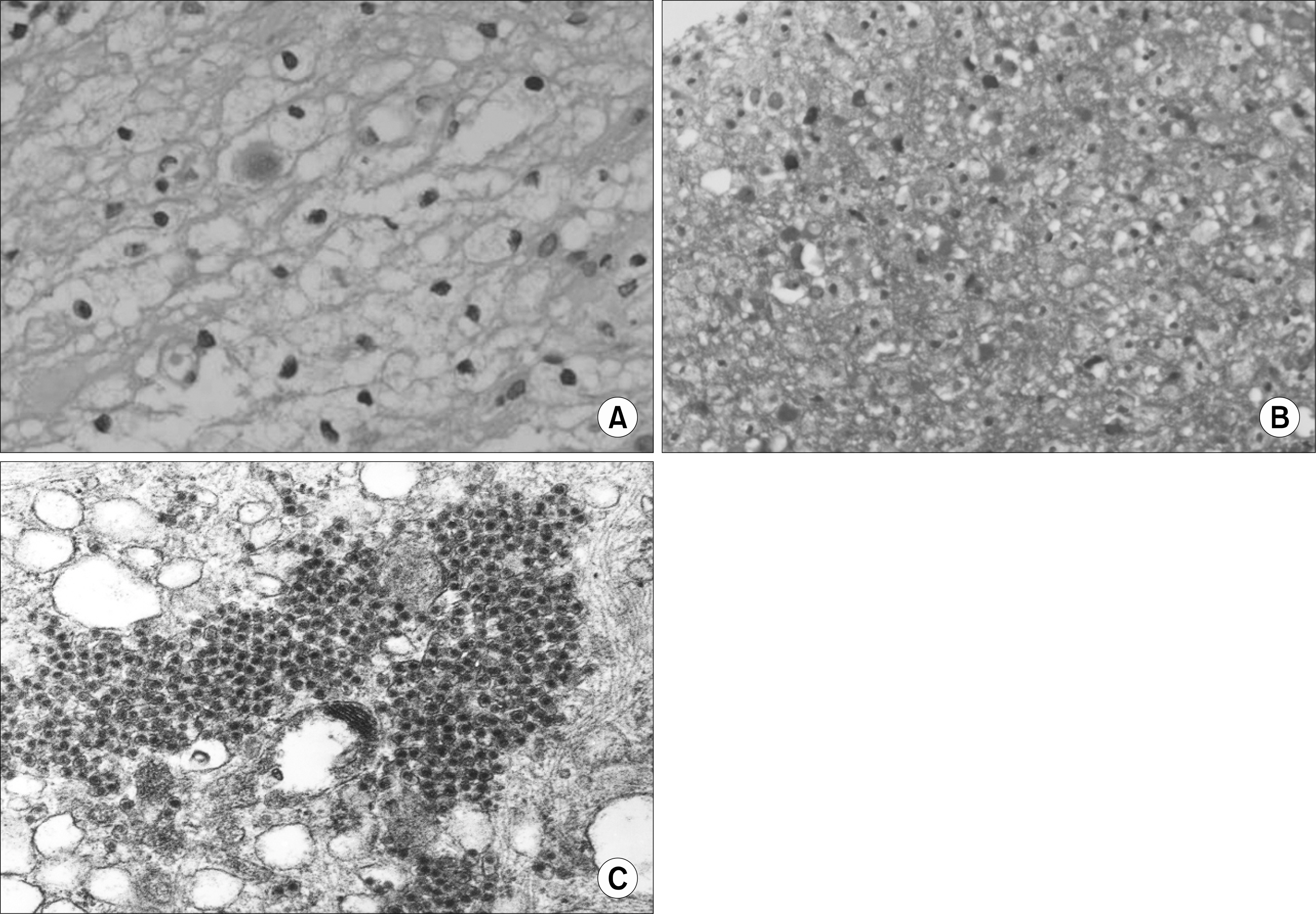
Fig. 2. Sections of brain tissue. (A) Hematoxylin and eosin stained section show some atypical oligodendrocytes with infiltraion of lymphocytes and foamy macrophages (×400, H&E). (B) Luxol fast blue stained section show destroyed myelin fiber (×200, Luxol Fast Blue). (C) Electron Microscopic finding show 33 nm-sized viral particles (×76,000).
Cited by 2 articles
-
A Case of Progressive Multifocal Leukoencephalopathy in Acquired Immune Deficiency Syndrome Initially Presented with Early Onset Dementia
Pyeong Kang Park, Jung Geun Oh, Seong-Ho Koh, Kyu-Yong Lee, Young Joo Lee, Hojin Choi
Dement Neurocogn Disord. 2014;13(1):20-23. doi: 10.12779/dnd.2014.13.1.20.A Case of Progressive Multifocal Leukoencephalopathy in Acquired Immune Deficiency Syndrome Initially Presented with Early Onset Dementia
Pyeong Kang Park, Jung Geun Oh, Seong-Ho Koh, Kyu-Yong Lee, Young Joo Lee, Hojin Choi
Dement Neurocogn Disord. 2014;13(1):20-23. doi: 10.12779/dnd.2014.13.1.20.
Reference
-
References
1. Greenlee JE. Progressive multifocal leukoencephalopathy–progress made and lessons relearned. N Engl J Med. 1998; 338:1378–80.2. Calabrese LH, Molloy ES, Huang D, Ransohoff RM. Progressive multifocal leukoencephalopathy in rheumatic diseases: evolving clinical and pathologic patterns of disease. Arthritis Rheum. 2007; 56:2116–28.
Article3. Warnatz K, Peter HH, Schumacher M, Wiese L, Prasse A, Petschner F, et al. Infectious CNS disease as a differential diagnosis in systemic rheumatic diseases: three case reports and a review of the literature. Ann Rheum Dis. 2003; 62:50–7.
Article4. 심민근, 김조헌, 박창수, 김형석, 최유덕, 이민철. 면 역 저하 환자들에게서 발생한 진행다초점백색질 뇌증 – 3예 보고 −. 대한병리학회지. 2007; 41:358–61.5. Brey RL. Neuropsychiatric lupus: clinical and imaging aspects. Bull NYU Hosp Jt Dis. 2007; 65:194–9.6. Haider S, Nafziger D, Gutierrez JA, Brar I, Mateo N, Fogle J. Progressive multifocal leukoencephalopathy and idiopathic CD4+lymphocytopenia: a case report and review of reported cases. Clin Infect Dis. 2000; 31:20–2. E.
Article7. Kokubun N, Ishihara T, Nishibayashi M, Ikeda S, Nagashima K, Hirata K. Progressive multifocal leukoencephalopathy with idiopathic CD4 positive T-lymphocytepenia mimicking a low grade glioma on proton MR spectroscopy. A case report. Rinsho Shinkeigaku. 2005; 45:663–8.8. Chikezie PU, Greenberg AL. Idiopathic CD4+ T lymphocytopenia presenting as progressive multifocal leukoencephalopathy: case report. Clin Infect Dis. 1997; 24:526–7.
Article9. Iwase T, Ojika K, Katada E, Mitake S, Nakazawa H, Matsukawa N, et al. An unusual course of progressive multifocal leukoencephalopathy in a patient with idiopathic CD4+ T lymphocytopenia. J Neurol Neurosurg Psychiatry. 1998; 64:788–91.
Article10. Garrels K, Kucharczyk W, Wortzman G, Shandling M. Progressive multifocal leukoencephalopathy: clinical and MR response to treatment. AJNR Am J Neuroradiol. 1996; 17:597–600.11. Post MJ, Yiannoutsos C, Simpson D, Booss J, Clifford DB, Cohen B, et al. Progressive multifocal leukoencephalopathy in AIDS: are there any MR findings useful to patient management and predictive of patient survival? AIDS Clinical Trials Group, 243 Team. AJNR Am J Neuroradiol. 1999; 20:1896–906.12. Fazakerley JK, Buchmeier MJ. Pathogenesis of virus-induced demyelination. Adv Virus Res. 1993; 42:249–324.
Article13. Andrei G, Snoeck R, Vandeputte M, De Clercq E. Activities of various compounds against murine and primate polyomaviruses. Antimicrob Agents Chemother. 1997; 41:587–93.
Article14. Elphick GF, Querbes W, Jordan JA, Gee GV, Eash S, Manley K, et al. The human polyomavirus, JCV, uses serotonin receptors to infect cells. Science. 2004; 306:1380–3.
Article15. Owczarczyk K, Hilker R, Brunn A, Hallek M, Rubbert A. Progressive multifocal leucoencephalo-pathy in a patient with sarcoidosis–successful treatment with cidofovir and mirtazapine. Rheumatology (Oxford). 2007; 46:888–90.
Article
- Full Text Links
-
- Actions
-
Cited
- CITED
-
- Close
- Share
-
- Similar articles
-
- Progressive Multifocal Leukoencephalopathy in a Patient with Systemic Lupus Erythematosus
- Recurrent Complex Partial Seizures in a Patient with Progressive Multifocal Leukoencephalopathy
- A Case of Transverse Myelitis as a First Manifestation of Systemic Lupus Erythematosus
- Hypointense Rim on Susceptibility-Weighted Imaging in a Patient with Progressive Multifocal Leukoencephalopathy
- A Ruptured Aneurysm in a Patient with Systemic Lupus Erythematosus: Case Report
